
Оставить заявку
Для заказа и получения более подробной информации оставьте заявку, наш менеджер свяжется с Вами!
Нажимая на кнопку, вы даете согласие на обработку персональных данных и соглашаетесь c политикой конфиденциальности
БОРЬБА С МИКРОБНЫМИ УГРОЗАМИ В СЕЛЬСКОМ ХОЗЯЙСТВЕ
с помощью интегративных методов визуализации и вычислительных подходов
Никхил Кумар Сингх, Аник Дутта, Гвидо Пуччетти, Дэниел Кролл
a Лаборатория эволюционной генетики, Институт биологии, Невшательский университет, CH-2000 Невшатель, Швейцария
b Фитопатология, Институт интегративной биологии, ETH Zürich, CH-8092 Цюрих, Швейцария
c Syngenta Crop Protection AG, CH-4332 Штайн, Швейцария
Автор для корреспонденции: daniel.croll@unine.ch (Д. Кролл)
DOI: 10.1016/j.csbj.2020.12.018. Получено 1 сентября 2020 г.; исправленная версия — 8 декабря 2020 г.; принято 13 декабря 2020 г.; опубликовано онлайн 29 декабря 2020 г.
Ключевые слова: сельское хозяйство; фитопатогены; высокопроизводительное фенотипирование; анализ изображений; полногеномное картирование ассоциаций; устойчивое развитие.
Аннотация
Патогены и вредители являются одной из основных угроз продуктивности сельского хозяйства во всем мире. На протяжении десятилетий целенаправленная селекция устойчивости использовалась для создания сортов сельскохозяйственных культур, устойчивых к патогенам и стрессу окружающей среды, сохраняя при этом урожайность. Процесс скрещивания, селекции и полевых испытаний для создания нового сорта, который зачастую длится десятилетия, сталкивается с проблемой быстрого роста количества патогенов, преодолевающих устойчивость. Аналогичным образом, противомикробные соединения могут быстро потерять эффективность из-за развития резистентности. Здесь мы рассматриваем три основные области, в которых вычислительные, визуальные и экспериментальные подходы совершают революцию в борьбе с повреждением сельскохозяйственных культур патогенами. Распознавание и оценка болезней растений значительно улучшились благодаря высокопроизводительным методам визуализации, применимым как в хорошо контролируемых тепличных условиях, так и непосредственно в полевых условиях. Однако компьютерное видение сложных фенотипов заболеваний потребует значительных улучшений. Параллельно с этим экспериментальные установки, подобные высокопроизводительным скринингам поиска лекарств, позволяют проверять тысячи штаммов патогенов на предмет изменений в резистентности и других соответствующих фенотипических признаков. Конфокальная микроскопия и флуоресценция позволяют получить богатую фенотипическую информацию о генотипах патогенов. Благодаря подходам картирования ассоциаций по всему геному, фенотипические данные помогают разгадать генетическую архитектуру признаков, связанных со стрессом и вирулентностью, ускоряя селекцию устойчивости. Наконец, совместный крупномасштабный скрининг изменчивости признаков сельскохозяйственных культур и патогенов может дать фундаментальное представление о том, как патогены сталкиваются с компромиссами при адаптации к устойчивым сортам сельскохозяйственных культур. Мы обсуждаем, как будущее внедрение таких инновационных подходов в селекции и скрининге патогенов может привести к более надежному контролю над болезнями. Ó 2020 Автор(ы). Опубликовано Elsevier B.V. от имени Исследовательской сети вычислительной и структурной биотехнологии. Это статья в открытом доступе по лицензии CC BY (http://creativecommons.org/licenses/by/4.0/).
Содержание
1. Введение
1.1. Знание врага: как патогены взаимодействуют с растениями, вызывая болезни
1.2. Высокопроизводительная оценка здоровья растений в полевых условиях
1.3. Проксимальное фенотипирование растений
1.4. Скрининг патогенов для повышения долговечности устойчивости растений
1.5. Расшифровка сложных взаимодействий «хозяин–патоген»: дальнейший путь
1.6. Использование слабостей патогенов, обусловленных компромиссами
2. Резюме и перспективы
1. Введение
Чтобы накормить население мира, необходимо стабильное производство безопасных продуктов питания, которому угрожают такие факторы, как изменение климата, деградация земель и болезни. Патогены и вредители вызывают значительное снижение продуктивности сельского хозяйства во всем мире, а стратегии борьбы со многими патогенами остаются неэффективными [1–3]. Около 20–30% мирового урожая теряется из-за болезней растений, а дополнительные послеуборочные потери достигают 12% [4]. Двумя основными подходами к защите сельскохозяйственных культур от болезней являются выведение устойчивых сортов, часто несущих определенные локусы, придающие устойчивость (т.е. R-гены) [5] и применение химических соединений (например, фунгицидов, инсектицидов) [6–8]. Целенаправленная селекция устойчивости практикуется на протяжении десятилетий для создания сортов, устойчивых к патогенам, но сохраняющих желаемые сельскохозяйственные характеристики. Тем не менее, даже современные методы молекулярной селекции часто включают в себя многолетние скрещивания, отбора и испытаний для создания нового сорта [9].
В то же время фитопатогены часто быстро преодолевают иммунитет, опосредованный генами устойчивости, срывая дорогостоящие усилия по селекции [10,11]. Аналогичным образом, внедрение синтетических химикатов часто сопровождается ростом популяций устойчивых патогенов [12,13]. Борьба с рисками продовольственной безопасности, создаваемыми патогенами, потребует объединения фундаментальных знаний о биологии болезней, достижений в высокопроизводительных подходах и инноваций в вычислительных инструментах. Практика устойчивого управления сельским хозяйством во многом зависит от понимания биологии и эволюционного потенциала основных патогенов сельскохозяйственных культур. Возникающие или повторно возникающие патогены можно сдержать только путем исследования их происхождения и путей миграции, а также их потенциала контрадаптации к принятым мерам контроля. Достижения в области геномики, транскриптомики и протеомики сыграли важную роль в выяснении этих вопросов и раскрыли широкий спектр молекулярных механизмов, регулирующих взаимодействия хозяина и патогена, что привело к более эффективным стратегиям контроля [14–17].
Редактирование генов и селекция, ориентированная на использование естественной генетической изменчивости, предоставляют важнейшие ресурсы для внедрения новых аллелей в усилия по улучшению сельскохозяйственных культур [18,19]. Генетическое улучшение сельскохозяйственных культур требует крупномасштабного скрининга тысяч линий, выращенных в различных условиях окружающей среды [20,21]. Что касается патогенов, первостепенное значение имеет быстрое выявление возникающей устойчивости к химическим веществам или вирулентности ранее устойчивых культур [3,22]. Генотипирование растений и патогенов достигло впечатляющей производительности при низких затратах, однако эквивалентные улучшения в высокопроизводительном скрининге фенотипической информации в значительной степени отстают [20,23]. Это, в свою очередь, создало препятствия для усилий по скоростной селекции и автоматизированному мониторингу состояния здоровья растений во время сельскохозяйственного производства.
Чтобы подчеркнуть недавние разработки, которые могут исправить эти недостатки, мы сначала обсудим важнейшие компоненты взаимодействия хозяина и патогена, на которые можно воздействовать. Затем мы рассмотрим недавние разработки в области феномики растений, ориентированных на болезни, ставшие возможными благодаря высокопроизводительным платформам фенотипирования. После этого мы подчеркиваем достижения в скрининге популяций патогенов для выявления ранних признаков эволюции резистентности или повышенной вирулентности.
Мы утверждаем, что объединение вышеупомянутых подходов позволило получить впечатляющее представление о генетической архитектуре болезней сельскохозяйственных культур. Наконец, мы обсудим, как инновационные подходы могут привести к более надежному контролю заболеваний.
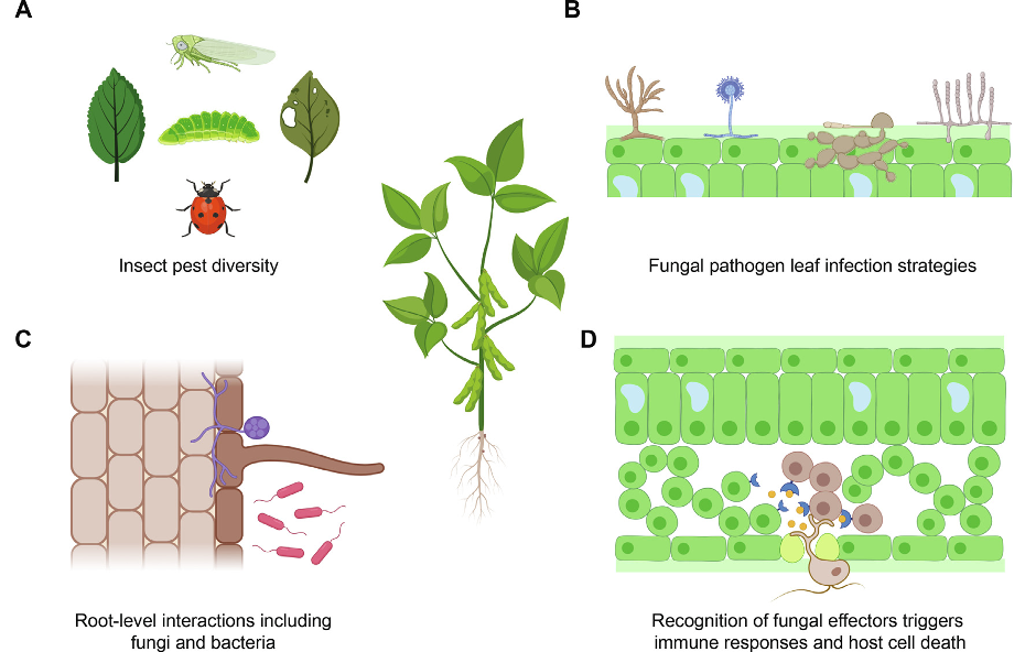
Рис. 1. Биология взаимодействия растений с патогенами. А) Листьями питаются многие насекомые, будучи травоядными. Б) Продольный разрез корня и ризосферы. Подземные взаимодействия могут оказывать существенное влияние на фенотип стебля и на все растение. Нитчатые грибы и бактерии могут оказывать благотворное воздействие на растение, облегчая усвоение фосфора и азота, или губительно действовать при столкновении с корневыми возбудителями. В) Дорзивентральный срез листа, колонизированный различными патогенными нитчатыми грибами. Слева: грибы родов Fusarium, Aspergillus, Magnaporthe и Podosphaera. Г) Нитчатые грибы и оомицеты, вызывающие инфекции листьев. Нитчатые возбудители могут проникать в мезофилл через устьица. Попав внутрь мезофилла, патогены колонизируют всю ткань в течение нескольких дней или недель. В зависимости от образа жизни (биотрофный или некротрофный) патогены выделяют небольшие белки (называемые эффекторами) для манипулирования растительными клетками. Растения, обнаруживающие эффекторы (синие рецепторы), могут вызвать сверхчувствительную реакцию (HR), приводящую к аутофагической гибели клеток, предотвращающей распространение патогена. (Для интерпретации ссылок на цвет в легенде к рисунку читатель отсылается к веб-версии этой статьи.)
1.1. Знание врага: как патогены взаимодействуют с растениями, вызывая болезни
Основные болезни сельскохозяйственных культур вызываются патогенами, включая грибы, бактерии и вирусы (рис. 1). Кроме того, насекомые-вредители также наносят существенные потери сельскохозяйственному производству [24,25]. Споры грибов и бактерий могут передаваться ветром, брызгами дождя и животными, включая человека и насекомых [26,27]. Некоторые патогены проникают в ткани растений (рис. 1Б) с помощью векторов и затем окружаются цитоплазмой, клеточной мембраной или клеточными стенками [28,29]. В других случаях возбудитель контактирует с внешней поверхностью растения, а затем задействует механизм проникновения, который может включать узкоспециализированные структуры (например, аппрессории) [30]. После инвазии в ткань хозяина успех патогена на конкретном хозяине во многом объясняется взаимодействием генных продуктов, закодированных в геномах хозяина и патогена [31,32]. Гены устойчивости (R) в растениях кодируют белки, которые прямо или косвенно обнаруживают наличие эффекторов патогена (также известных как факторы авирулентности или Avr) и вызывают сильный иммунный ответ (рис. 1B-D).
Патогены могут избежать распознавания за счет диверсификации последовательностей или удаления эффекторных генов [33]. Несмотря на важность отдельных локусов, контролирующих исход инфекции, патогенность может иметь сложную и в значительной степени количественную генетическую основу [34–39]. Для успешного заражения растений патогенам также необходимо выдерживать ряд абиотических стрессовых факторов (рис. 1В). Поскольку виды патогенов имеют оптимальный диапазон условий окружающей среды (т.е. температуры, pH, влажности) для процветания, изменения в окружающей среде могут изменить способность патогена причинять вред [40]. Факторы окружающей среды, такие как годовые колебания температуры, количество и характер осадков, уровни CO2 и озона, могут влиять на тяжесть заболеваний растений [2,41]. Кроме того, эффективность пестицидов может быть существенно изменена погодными условиями (например, смывом фунгицидов после сильных дождей). Следовательно, задача сдерживания ущерба от патогенов в сельском хозяйстве состоит в том, чтобы предсказать появление вирулентных штаммов и рост устойчивости к фунгицидам.
Учитывая возможные серьезные последствия, решающее значение имеет раннее выявление снижения устойчивости сельскохозяйственных культур или потери чувствительности к пестицидам. Обнаружение в начале вегетационного периода и идентификация ранее не охарактеризованных патогенов остаются серьезными проблемами. Классическая диагностика болезней растений обычно основана на визуальной оценке симптомов обученными людьми, классифицирующими тяжесть заболевания по линейным шкалам [42]. Однако классическая диагностика, очевидно, возможна только при появлении симптомов [43]. Некоторые патогены (например, биотрофы) могут питаться питательными веществами растений в течение длительного периода, не вызывая явных симптомов инфекции [44]. Патогены, убивающие растительные клетки для получения питательных веществ, обнаружить более очевидно [45]. Усложняет выявление патогенов и то, что многие патогены могут претерпевать временные изменения в инфекционном образе жизни [46].
Заражение растений часто происходит на участках с большими площадями полей, свободными от болезней на ранней стадии заражения. Это связано с распространением возбудителей на короткие расстояния вокруг исходных очагов заболевания [47,48]. Следовательно, необходима эффективная и крупномасштабная оценка заболеваемости для выявления инфекций на ранних стадиях, еще до того, как симптомы станут видимыми.
Дистанционная визуализация и расширенный анализ данных могут использоваться для выявления очагов заболеваний и обоснования разумного применения химических средств борьбы (рис. 2). Сельскохозяйственная среда представляет собой множество сложностей, которые часто плохо отражаются в тепличных экспериментах. На полях растения сталкиваются с меняющимися условиями окружающей среды и конкурируют за свет, воду и питательные вещества. Следовательно, высокопроизводительные инструменты фенотипирования и анализа в основном подразделяются на подходы, применимые в полевых условиях, и лабораторные/тепличные установки. Симптомы заболевания могут включать в себя любой диапазон изменений цвета, формы или функционирования растения по мере его реакции на патоген и могут визуализироваться при определенных длинах волн (рис. 2А). В зависимости от возбудителя симптомы заболевания могут варьироваться от пятен листьев, хлороза, некроза, увядания или чрезмерного разрастания [49,50].
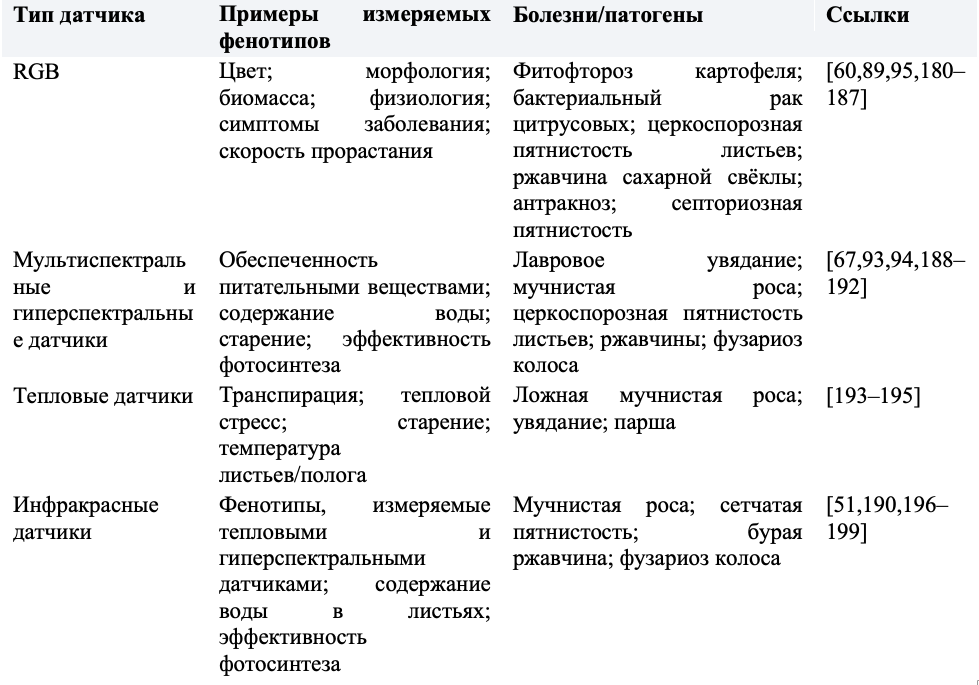
Однако стресс растений, помимо инфекций, может активировать защитные механизмы, приводящие к субоптимальному росту, потере хлорофилла или изменениям температуры поверхности [51,52]. Такие изменения вызывают заметные сдвиги в спектральной характеристике по сравнению со здоровым растением и могут быть измерены различными методами (рис. 2A-B, таблица 1) [53]. Системы визуализации полагаются на количественную оценку характеристик поглощения, пропускания или отражения электромагнитного излучения, взаимодействующего с поверхностью растения (рис. 2А-Б) [54]. Мы предоставляем набор конкретных фенотипов, связанных с заболеванием, которые могут быть зафиксированы различными датчиками камеры по всему спектру длин волн в таблице 1. Например, в коротковолновом инфракрасном спектре (1000–3000 нм) вода и биомолекулы демонстрируют характерные закономерности отражения (рис. 2А). Коротковолновый инфракрасный датчик может обнаружить зараженную область на основе различий в содержании воды, вызванных болезнью (рис. 2Б).
Затем вычислительные алгоритмы могут выявить контрасты между зараженными и здоровыми участками, чтобы оценить тяжесть заболевания на уровне листьев (рис. 2B). Различные алгоритмы, доступные для оценки фенотипа растений, недавно были рассмотрены более подробно [55]. Оценка фенотипа заболевания также во многом зависит от экспериментальной установки. Фенотипирование болезней растений в условиях теплицы или ростовой камеры в значительной степени устраняет колебания, связанные с окружающей средой, и эксперименты можно легко повторить для тех же генотипов. Такие эксперименты с конечной точкой также позволяют инвазивную оценку колонизации патогенов на поверхности и внутри тканей растений с использованием поперечной визуализации целых органов (например, листьев, рис. 2C), широкопольной и конфокальной микроскопии (рис. 2C), а также различных полимеразных цепных реакций (ПЦР) и методов, основанных на секвенировании, для оценки численности. Такие хорошо контролируемые измерения, полученные в основном в тепличных экспериментах, использовались для картирования QTL и полногеномных исследований ассоциаций (GWAS) для обнаружения маркеров, связанных с устойчивостью, и информирования программ селекции.
1.2. Высокопроизводительная оценка здоровья растений в полевых условиях
Широкое распространение получили технологии, подходящие для скрининга болезней растений даже на ранних стадиях заражения [20]. Платформы фенотипирования, объединяющие такую технологию, позволяют осуществлять как проксимальный, так и удаленный мониторинг отдельных листьев растений (или других органов), отдельных растений, а также целых полей (рис. 2C). Регистрация фенотипов растений в полевых условиях состоит либо из измерений из космоса, либо с воздуха с помощью камер, установленных на спутниках (WorldView-3, www.digitalglobe.com), беспилотных летательных аппаратах (БПЛА), парашютах, дирижаблях, пилотируемых ротокоптерах или системах с неподвижным крылом [20,56,57]. Спутниковая съемка может обеспечить многоспектральные изображения с разрешением от нескольких метров до сотен метров [57]. Однако существуют серьезные ограничения, связанные с погодными условиями, частотой съемки изображений и общими затратами. Спутниковые изображения также наиболее полезны для оценки растительного покрова в региональном или континентальном масштабе, а не для испытаний селекции сельскохозяйственных культур на отдельных полях.
Недавние улучшения в спутниковых изображениях высокого разрешения, например GF-1 из Китая или SPOT из Европы могут обеспечить фенотипирование отдельных полей с временным разрешением в метровом масштабе [58,59]. Такое разрешение позволяет уловить изменения отражательной способности, вызванные старением или вспышками патогенов. Беспилотные летательные аппараты (БПЛА) стали популярными в последние годы из-за простоты развертывания и возможности установки систем захвата изображений высокого разрешения [56,60]. На БПЛА может быть установлен ряд датчиков в зависимости от грузоподъемности и типа требуемых данных. В зависимости от спектра и количества полос датчики можно разделить на несколько типов, включая видимый свет (RGB) [61–63], мультиспектральные, гиперспектральные [64–67], тепловые инфракрасные [68,69] и трехмерные лазерные изображения [70,71]. В качестве примера применения можно назвать коммерческий квадрокоптерный БПЛА, оснащенный цифровыми и мультиспектральными камерами, который позволил обнаруживать и контролировать рис.
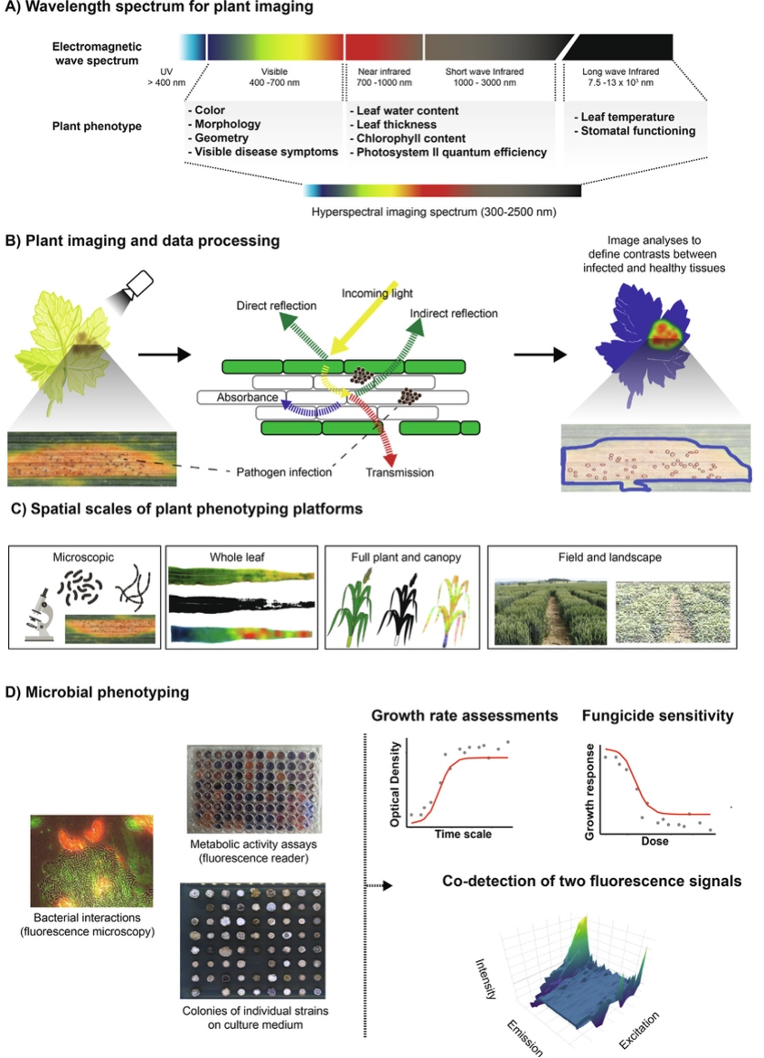
Рис. 2. Методы высокопроизводительного фенотипирования растений и патогенов. А) Свет видимого спектра можно использовать для обнаружения изменений цвета и морфологии зараженной растительной ткани. Инфракрасное и коротковолновое инфракрасное излучение позволяют регистрировать изменения содержания воды, толщины листьев и эффективности фотосинтеза. Длинноволновое инфракрасное излучение позволяет оценивать температуру поверхности растений. Гиперспектральные датчики захватывают несколько изображений в диапазоне 300–2500 нм. Б) Системы визуализации оценивают характеристики поглощения, передачи или отражения падающего электромагнитного излучения, взаимодействующего с поверхностью растения. Ткань больного растения часто отличается по отражательной способности от здоровой ткани. Алгоритмы анализа изображений определяют контрасты между больными и здоровыми участками листьев. В) Пространственные масштабы подходов к фенотипированию растений. Г) Скрининг популяций патогенов можно проводить в жидких культурах или на твердых средах. В наиболее распространенных экспериментах отслеживают темпы роста путем оценки плотности культуры с течением времени или оценки кривых «доза-реакция» при воздействии противомикробных соединений. Совместные культуры нескольких микробов можно анализировать с использованием двух отдельных пар испускания/возбуждения, специфичных для каждого совместно культивируемого вида. гниение оболочек [66]. Гексакоптер, оснащенный RGB-камерами, позволил измерить высоту растений пшеницы, что соответствовало измерениям вручную и наземным датчиком LiDAR [72]. Отслеживать развитие фитофтороза на картофельном поле можно было с помощью воздушной RGB-камеры [60]. Тепловые инфракрасные датчики также успешно использовались для количественной оценки реакции растений на стресс засухи у черного тополя [73], почвенного покрова сорго, температуры кроны сахарного тростника и полегания посевов пшеницы [74]. Основным ограничением фенотипирования на основе аэрофотокамеры являются ошибки, возникающие при создании ортофотопланов, которые представляют собой составные изображения сшитых и геометрически скорректированных фотографий [57,75]. Неточности в ортофотопланах могут быть вызваны низким разрешением сенсора камеры, высотой и тем, как определялось положение камеры во время съемки [76]. Однако усовершенствования в алгоритмах исправления и технологиях дронов значительно уменьшили ошибки ортофотоплана [77]. Неточности ортомозаики также можно исправить, дополняя изображения, полученные с помощью проксимальных платформ фенотипирования [78,79].
1.3. Проксимальное фенотипирование растений
Проксимальное фенотипирование чаще всего применяется в тепличных экспериментах или в хорошо контролируемых полевых экспериментах. Датчики, охватывающие широкий диапазон спектров, можно устанавливать на стационарных платформах или, при использовании вне помещений, на подвесных тросах, роботах и тракторах [20,23,80–84]. Фиксированная структура на большом поле имеет преимущества перед фенотипированием как с помощью транспортных средств, так и с помощью БПЛА, и не ограничивается полезной нагрузкой датчиков и емкостью батареи [85]. Например, платформа полевого фенотипирования (FIP), созданная в ETH Zurich, использует подвесную кабельную установку для перемещения датчиков по большой экспериментальной полевой площадке [84]. Подобные установки существуют в Университете Небраски для фенотипирования культур кукурузы и сои [86]. Эти платформы проксимального фенотипирования выигрывают от наличия нескольких установленных датчиков, собирающих информацию о фенотипических признаках в широком диапазоне длин волн.
Например, проксимальная гиперспектральная сенсорная платформа была способна обнаружить заражение мучнистой росой только на основе исследований полога ячменя [67]. Визуализация, основанная на флуоресценции, часто используется для проксимального фенотипирования для количественной оценки фенотипических изменений, связанных с фотосинтезом, содержанием азота и такими заболеваниями, как пятнистость Septoria tritici (рис. 2A-B) [42,87–89]. Платформы проксимального фенотипирования могут измерять температуру кроны, содержание азота, а также фенотипические признаки, включая площадь листьев и высоту растений [90]. Такое эффективное фенотипирование всего растения помогло ускорить отбор устойчивых к засухе растений риса [89]. Платформа была разработана для визуализации в видимом спектре для регистрации морфологических особенностей. Система также включала инфракрасные и ближние инфракрасные изображения для количественного определения содержания воды, а также измерения температуры и флуоресценции для количественной оценки эффективности фотосинтеза [89].
Платформа могла точно различать толерантные и восприимчивые растения, что позволяло осуществлять быстрый отбор для скоростной селекции. Последующие приложения включали функциональные генетические исследования устойчивости к засухе и приложения GWAS, которые, в свою очередь, могут способствовать усилиям по селекции с использованием маркеров. В другом применении флуоресценция хлорофилла и термическое (инфракрасное) фенотипирование около 300 сортов иранской пшеницы в сочетании с GWASвыявили адаптивные аллели к стрессу засухи, полезные для маркерной селекции [91]. Современные технологии фенотипирования растений и алгоритмы обработки изображений с трудом позволяют надежно дифференцировать симптомы заболеваний, вызванных множественными или неизвестными патогенами [92,93]. Частично это связано со сложностью симптомов грибковых и бактериальных заболеваний сельскохозяйственных культур, но также и с тем, что симптомы заболевания на ранних стадиях заражения вряд ли являются диагностическими для конкретных патогенов.
Дефицит питательных веществ или воды (при отсутствии патогенов) может вызывать симптомы, которые трудно отличить от фенотипов заболевания. Методы молекулярного обнаружения (например, qPCR) часто необходимы для четкого установления того, что может вызвать заболевание. Интересными разработками для преодоления этих ограничений являются, например. спектры дифференциального отражения (400–1050 нм) листьев сахарной свеклы, которые помогают различать три различных грибковых патогена, включая Cercospora beticola, Erysiphebetae и Uromyces betae [94]. Комбинированные данные теплового изображения и изображения в видимом свете были переданы в алгоритм машинного обучения для дифференциации инфекций, вызванных грибком мучнистой росы томата Oidium neolycopersici [95].
Одним из основных ограничений высокопроизводительного фенотипирования болезней растений являются временные рамки обнаружения болезней. Визуализация, основанная на снижении фотосинтеза как индикаторе прогрессирования заболевания, может обнаружить симптомы только после того, как патоген проникает в ткани растения и снижает активность хлорофилла (рис. 1, 2). Гиперспектральные или мультиспектральные датчики позволяют отличить больные листья от здоровых гораздо раньше, чем визуализация только в видимом спектре. Абдулрида и др. [96] смогли обнаружить и различить возникновение целевой пятнистости, вызванной грибом Corynespora cassicola, и бактериальной пятнистости, вызванной Xanthomonas perforans, на томатах в бессимптомной фазе. Подход к мультиспектральной визуализации оказался успешным как в лабораторных, так и в полевых условиях. Такие надежные методы фенотипирования могли бы позволить обнаружить очаги инфекции в полевых условиях. Возникающие инфекции затем можно было бы изолировать и лечить индивидуально, применяя специфические фунгициды.
Технологии фенотипирования растений позволили получить данные для заполнения общедоступных баз данных изображений (подведены итоги в [97]). Такие данные могут быть использованы для машинного обучения и показывают многообещающие результаты в прогнозировании возникновения заболеваний и идентификации видов возбудителей [95,98,99]. Используя общедоступный набор данных из 54 306 изображений здоровых и больных листьев растений, сеть глубокого обучения смогла выявить 26 различных заболеваний с точностью 99,35% [99]. Развитие машинного обучения применительно к проблеме фенотипирования растений ограничивалось в основном двумя факторами: i) ограничением публичного доступа к информации.
Таблица 1. Оценка заболеваний с использованием высокопроизводительных датчиков.
Таблица 1. Оценка заболеваний с использованием высокопроизводительных датчиков.
Ограниченность общедоступных баз данных изображений фенотипов болезней сельскохозяйственных культур в различных условиях окружающей среды и изображений фенотипов болезней сельскохозяйственных культур в различных условиях окружающей среды и ii) плохих аннотаций данных изображений и зафиксированных симптомов [100,101]. Отсутствие надежных обучающих наборов данных вынудило исследователей использовать неоптимальные базы данных, что привело к неоднозначности в различении фенотипов заболеваний от старения или других факторов окружающей среды [99,100,102]. Таким образом, несмотря на огромный потенциал машинного обучения для оценки болезней растений, текущие реализации необходимо будет улучшить с помощью больших наборов справочных данных.
1.4. Скрининг патогенов для повышения долговечности устойчивости растений
Взаимодействия растений и микробов часто являются предметом быстрой коэволюционной динамики, изменяющей генетический состав как хозяина, так и патогена [103]. Эволюционирующие патогены делают селекцию устойчивости явно сложной задачей. Кроме того, значительно увеличились вспышки новых патогенов [104], в том числе, например. недавние вспышки Magnaporthe oryzae и Puccinia graminis f. сп. tritici в Бангладеш и Италии соответственно ставят под угрозу производство пшеницы [104–107]. Быстрое изменение популяций патогенов сельскохозяйственных культур наиболее очевидно через повышение устойчивости к фунгицидам [108,109], но многие другие фенотипы также демонстрируют высокую изменчивость [110,111]. Некоторые патогены приобрели свойства вирулентности путем приобретения генетического материала путем горизонтального переноса. Ярким примером является повышение вирулентности пшеницы за счет переноса ключевого гена, кодирующего токсин, между видами грибов [112].
Среди наиболее разнообразных возбудителей – космополитный гриб Zymoseptoria tritici, поражающий пшеницу. Устойчивость сортов в значительной степени была преодолена, и в течение нескольких лет возникла новая вирулентность [35,113]. Популяции Z. tritici генетически и фенотипически очень разнообразны, причем внутриполевое разнообразие приближается к уровням разнообразия в континентальном или глобальном масштабе [114–116]. Чтобы противостоять очень разнообразным и быстро развивающимся патогенам, программы селекции часто пытаются предотвратить снижение устойчивости путем объединения («пирамидирования») нескольких генов устойчивости [35,117]. Несмотря на проблемы, связанные с быстро развивающимися видами патогенов, наследственную изменчивость признаков можно использовать для выявления механизмов, лежащих в основе вирулентности [38,118]. Ряд основных патогенов сельскохозяйственных культур легко культивируются в стерильных лабораторных условиях, что позволяет воспроизводить оценку экспрессии признаков.
Клональное размножение в стерильной среде позволяет эффективно реплицировать фенотипы, экспрессируемые разными генотипами. Основным применением высокопроизводительного скрининга патогенов является измерение чувствительности к фунгицидам, что важно для раннего обнаружения мутаций устойчивости в региональном или континентальном масштабе [119]. В более широком смысле, оценка реакции грибковых и бактериальных патогенов на стресс может дать важные сведения об экспрессии факторов вирулентности, множественной лекарственной устойчивости, образовании биопленок и устойчивости к противомикробным препаратам [117,120]. Это связано с тем, что стресс, вызванный в условиях in vitro, может иметь сходство с состояниями инфекционного стресса. На стадиях инфекции патогенам приходится справляться с различными защитными реакциями хозяина, включая лишение питательных веществ, изменение pH и т. д. Следовательно, условия in vitro могут быть полезными триггерами для экспрессии белков, связанных с вирулентностью.
Помимо измерения скорости роста как показателя физиологического состояния, фенотипические скрининги могут быть расширены до измерения гетерогенности формы спор [121], жизнеспособности клеток как процента живых клеток или жизнеспособности клеток, определяемой как физиологические возможности клетки [122,123] и могут быть оценены с помощью проточной цитометрии и показателей флуоресценции. Более подробный анализ сосредоточен на вариациях жизненного цикла инфекции у хозяина [124]. Наконец, оценка температурной адаптации может дать важные сведения об адаптивном потенциале и способности патогенов справляться с климатом будущего [124,125].
Однако для повышения производительности ручные процедуры обработки необходимо будет заменить роботизацией. Платформы высокопроизводительного фенотипирования микробных организмов значительно продвинулись в автоматизации культивирования клеток, обработки жидкостей, нанесения пятен на культуральную среду в виде плотных массивов и интеграции измерений флуоресценции и микроскопии [126–129]. Такие технологии часто впервые применялись для скрининга активных соединений в программах разработки лекарств в фармацевтической промышленности [130]. В последние годы было разработано несколько методов высокопроизводительного скрининга in vitro для исследования генов толерантности к лекарствам и выяснения аспектов патогенности [128, 129]. Высокопроизводительные конвейеры выигрывают от робототехники, такой как Rotor-HDA, которая может дублировать и создавать резервные копии больших библиотек штаммов в твердых или жидких средах в формате 96–6144 изолятов на чашку.
Рост и чувствительность к фунгицидам можно оценить по оптической плотности (ОП), размеру колоний или флуоресценции жидких культур (рис. 1D) [131]. Более того, цвета культуры являются мощным фактором производства меланина или других вторичных метаболитов [132]. Другие высокопроизводительные платформы включают мультиплексную микрофлюидную культуру клеток, автоматизированную программируемую обработку жидкостей для возмущения клеток, количественную покадровую микроскопию и компьютерный анализ покадровых фильмов [133]. Такие платформы наиболее полезны для экспериментов с возмущениями (например, осмотическим шоком или воздействием лекарства) с флуоресцентными показаниями, чувствительными к изменениям экспрессии генов или субклеточной локализации. Основная цель технологии — ускорить открытие лекарств путем скрининга больших библиотек противомикробных соединений со скоростью тысяч соединений в неделю. Также предпринимаются усилия по разработке высокопроизводительных подходов для создания мутированных версий мишеней лекарств [128].
Высокопроизводительные методы скрининга в дальнейшем используются для характеристики метаболических, фармакокинетических и токсикологических данных о новых лекарствах и снижения затрат на разработку противомикробных соединений [129,134]. Во время разработки нового пестицида полевые испытания могут оказаться чрезвычайно дорогостоящими. Таким образом, высокопроизводительный скрининг in vitro различных популяций патогенов очень информативен в отношении возможной устойчивой устойчивости, что может предотвратить быстрое снижение эффективности в полевых условиях [135]. Обширная информация о вариациях фенотипических признаков у видов патогенов в сочетании с недорогим секвенированием может быть использована для GWAS [136]. С момента дебюта метода в 2005 году GWAS выявил сотни тысяч однонуклеотидных полиморфизмов (SNP) и структурных вариантов, связанных с тысячами различных фенотипов, в основном ориентированных на людей [136–138]. Популяции патогенов растений часто содержат как вирулентные, так и авирулентные штаммы.
Следовательно, благодаря надежному и надежному фенотипированию анализы GWAS могут идентифицировать гены, лежащие в основе конкретного повышения вирулентности. Картирования популяций из ~100 штаммов было достаточно для идентификации ключевых факторов вирулентности [139–141] и локусов устойчивости к фунгицидам у грибковых патогенов [135,139,140,142,143]. Исследования генетического картирования также могут помочь в процессе разработки новых фунгицидов путем раннего выявления генов «горячих точек» устойчивости [135].Критическим фактором для успешного применения GWAS является наличие высококачественных геномных ресурсов, представляющих генетическое разнообразие видов патогенов.
Недавние пангеномные анализы фитопатогенов заложили основу для такой работы [144].
1.5. Расшифровка сложных взаимодействий хозяина и патогена – путь вперед
Применение визуализации в сочетании с вычислительными методами позволило добиться огромного прогресса в выведении устойчивых культур и обнаружении появления новых угроз патогенов. Чтобы упростить задачу, большинство исследований до сих пор фокусировались либо на вариациях со стороны растений, либо со стороны патогенов. Однако природа взаимодействий хозяин-патоген может варьироваться в зависимости от пространства и времени [145,146]. Помимо таких межгенотипических взаимодействий, на исход инфекций влияют условия окружающей среды (влажность, температура, конкурирующие микробы и т. д.). Сложность частично возникает из-за сложных циклов заражения патогенами, начиная с начальных контакт с хозяином для передачи новым хозяевам [147,148]. Каждая стадия инфекции, вероятно, в некоторой степени регулируется отдельными генами. Таким образом, улучшение наших возможностей прогнозировать исходы инфекций при множестве различных взаимодействий между растениями и средами обитания является важнейшим шагом в повышении устойчивости сельскохозяйственных культур и борьбе со вспышками болезней в долгосрочной перспективе.
Интеграция различных наборов данных омики для построения биологических сетей (например, сетей совместной экспрессии генов или сетей межбелковых взаимодействий) стала мощным подходом к раскрытию генетических факторов, контролирующих биологические взаимодействия [149–151]. Помимо этого, инновационные применения GWAS могут помочь идентифицировать причинные гены путем одновременного изучения генотипов хозяина и патогена в матрице инфекции [152,153] (рис. 3). Применяя такой подход к взаимодействию Arabidopsisthaliana и Xanthomonas arboricola, Wang et al. [152] идентифицировали специфические гены количественной устойчивости к болезням у хозяина, которые эффективны только против определенного набора штаммов патогена. Изучение болезней сельскохозяйственных культур как матрицы инфекции может помочь идентифицировать геномные регионы, лежащие в основе коэволюции хозяина и патогена, и гены, ответственные за определенные фенотипы [154–156]. Однако на данный момент практически отсутствуют приложения в сельскохозяйственном контексте, поскольку такие подходы требуют экспериментальных исследований и являются дорогостоящими.
Усовершенствованные методы инокуляции патогенов, такие как анализ отдельных листьев [157–159] или анализ головы [160] в сочетании с автоматическим анализом изображений [161], позволят снизить экспериментальную нагрузку. Следовательно, сбор точных фенотипических данных из большой матрицы инфекций хозяев и патогенов становится все более доступным [157]. Помимо увеличения размеров выборки анализируемых генотипов хозяина и патогена, требуется особая осторожность, чтобы адекватно охватить генетическое разнообразие хозяина и патогена. Используя глобальный набор популяций основного патогена пшеницы Z. tritici, был идентифицирован большой набор локусов, связанных с вирулентностью и размножением патогена на разных хозяевах [38,162]. Большая матрица инфекций, состоящая из 98 штаммов Botrytis cinerea и 90 генотипов растений восьми видов, выявила высокополигенную архитектуру вирулентности патогена и специализацию хозяина [163]. Следовательно, одновременное расширение скрининга генетического разнообразия хозяина и патогена обеспечивает мощный подход к всестороннему картированию генетической архитектуры вирулентности и устойчивости.
Наиболее важные для изучения патогены
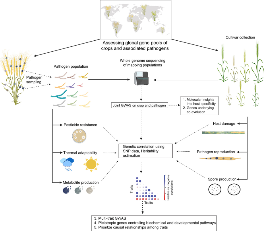
Рис. 3. Комплексная основа для определения генетической основы взаимодействия сельскохозяйственных культур и патогенов. Основу скрининга составляют генетически разнообразные популяции патогенов и сорта сельскохозяйственных культур из разных географических регионов. Секвенирование генома позволяет проводить совместные полногеномные ассоциативные исследования (GWAS) для определения генетической архитектуры вирулентности (у патогенов) и устойчивости (у сельскохозяйственных культур). Глобальные популяции как патогенов, так и сельскохозяйственных культур будут отражать наиболее важные генетические вариации. Помимо вирулентности, популяции патогенов можно проверять на наличие локусов, лежащих в основе устойчивости к пестицидам, термической адаптации и продукции метаболитов (например, меланина). Выявление генетических корреляций между признаками патогена облегчает идентификацию плейотропных генов, определяющих компромиссы. Некоторые иллюстрации предоставлены biorender.com в соответствии с условиями их использования. Тигате способны инфицировать несколько органов растения (например, черную ножку рапса) или на разных стадиях роста. Увеличение размера выборки как у хозяина, так и у патогена также улучшит оценку наследственности (например, как показано в исследованиях на людях [164]). В целом, расширение генетического разнообразия хозяев и патогенов при скрининге поможет выявить ранее неизвестные резистентности/восприимчивости в зародышевой плазме сельскохозяйственных культур.
1.6. Использование слабости патогенов, обусловленной компромиссами
То, насколько успешно патогены справляются с различными средами, зависит от компромиссов в жизненном цикле, которые возникают из-за дилемм распределения ресурсов и антагонистических действий генов. Компромисс указывает на то, что увеличение одного признака связано с уменьшением другого признака. Такие компромиссы обычно зависят от генетического фона хозяина и абиотических условий. Некоторые штаммы патогенов специализировались на определенных хозяевах или климатических условиях, чтобы максимизировать свою эффективность. Классические примеры включают дикую патосистему взаимодействия Plantago lanceolata и Podosphaera plantaginis [165,166]. Компромиссы, вероятно, являются ключевым фактором, поддерживающим полиморфизм в популяциях патогенов [167,168]. Исследования уже выявили компромисс между повреждением листьев и бесполой передачей [169], размером и количеством спорангиев [170], латентным периодом образования спор, размером и скоростью спорообразования [171].
Анализы чувствительных к фунгицидам и устойчивых к ингибиторам деметилирования (DMI) изолятов возбудителя сахарной свеклы C. beticola показали, что устойчивые изоляты имеют значительно более низкую вирулентность и образование спор, чем чувствительные изоляты [172]. Не обнаружено различий по инкубационным периодам, росту мицелия, прорастанию конидий и длине зародышевых трубок. Аналогично, у бактериального патогена Ralstonia solanacearum мутанты, лишенные гена синтеза экзополисахаридного фактора вирулентности, демонстрируют повышенную скорость роста по сравнению со штаммом дикого типа [173,174]. Кроме того, сельскохозяйственные патогены, вероятно, сталкиваются со многими дополнительными, пока неизвестными компромиссами, поскольку патогены также должны выживать за пределами однолетних культур-хозяев [175,176]. Генетическая основа компромиссов остается в значительной степени неизвестной, что препятствует использованию присущих патогенам слабостей.
Частично это связано с тем, что компромиссы обычно выражаются в корреляциях фенотипических признаков, которые часто смешиваются с вариациями окружающей среды и генетической субструктурой. Выявление генетических корреляций между признаками может обойти вышеупомянутые проблемы, поскольку оно отражает прямое влияние генетических факторов, контролирующих компромиссы, и устойчиво к искажающим факторам [177]. Недавнее исследование изучало генетические компромиссы, основанные на генетических корреляциях в патогене пшеничного грибка Z. tritici. Работа глобальной коллекции штаммов на двенадцати сортах пшеницы и в различных абиотических условиях выявила широкий плейотропный контроль активности патогенов на хозяине и вне его [162]. Слабость патогенов в конкретных средах будет способствовать более эффективному планированию борьбы с болезнями и предотвращению снижения резистентности в будущем. Следовательно, комплексные карты генетических компромиссов, возможно, позволят реализовать инновационные стратегии борьбы с болезнями (рис. 3).
Помимо выявления компромиссов, коррелирующие признаки можно комбинировать для выполнения мульти-признакового GWAS, чтобы точно определить плейотропные гены и определить причинно-следственную связь между признаками в конкретных средах [178,179] (рис. 3).
2. Резюме и перспективы
Технологический прогресс в оценке восприимчивости больших коллекций сельскохозяйственных растений к повреждению патогенами имеет решающее значение для современных усилий по селекции устойчивости. Разнообразные методы захвата изображений позволяют отслеживать повреждение растений на уровне клеток, листьев, всего растения или поля. Большинство приложений ориентированы на видимый спектр, но платформы гиперспектральной визуализации недавно получили возможность обнаруживать заражение патогенами еще до появления симптомов. Основной областью текущих исследований является улучшение алгоритмов анализа изображений для обнаружения и классификации повреждений, вызванных патогенами. Вариации внутри отдельных видов патогенов могут быть весьма информативными в отношении возникновения новой вирулентности или устойчивости к пестицидам. Робототехника, применяемая для автоматизации культивирования тысяч штаммов патогенов, позволяет проводить скрининг метаболических изменений, чувствительности к лекарствам и выработку вторичных метаболитов, улучшая наше понимание того, как патогены справляются с сельскохозяйственной средой.
Высокопроизводительное фенотипирование растений и патогенов можно сочетать с секвенированием генома и приложениями GWAS. Раскрытие генетической основы устойчивости хозяина помогает ускорить усилия по селекции посредством отбора с помощью маркеров. Анализ популяций патогенов может дать информацию о возможных компромиссах при возникновении вирулентности или устойчивости к пестицидам. Будущие направления исследований должны быть сосредоточены на ряде дополнительных областей исследований.
· Создать эффективные конвейеры, объединяющие методы визуализации и молекулярные анализы для обнаружения патогенов. Такие интегрированные системы могут помочь фермерам принять соответствующие контрмеры на полях до того, как будет нанесен масштабный ущерб, и сократить общее применение пестицидов.
· Восприимчивость сортов сельскохозяйственных культур к основным патогенам должна постоянно переоцениваться для выявления изменений в профиле вирулентности преобладающих патогенов. Быстрая эволюция патогенов может привести к катастрофическому нарушению устойчивости и должна быть обнаружена достаточно рано. Высокопроизводительные системы визуализации, фиксирующие симптомы заболеваний, должны сочетаться с машинным обучением для эффективного распознавания изменений в профилях вирулентности. Отсутствие курируемых и открытых баз данных изображений заболеваний в настоящее время замедляет прогресс.
· Усилия по региональному мониторингу снижения устойчивости или потери эффективности пестицидов могут быть достигнуты путем высокопроизводительного геномного скрининга зараженного листового материала. Эффективные анализы генотипирования, ориентированные на основные гены, вероятно, хорошо масштабируются для широкого применения. Биоинформационные процедуры для такого анализа геномных данных в основном существуют. Успешное внедрение такого геномного мониторинга также поможет обнаружить появление новых патогенов на достаточно раннем этапе, чтобы можно было вывести устойчивые сорта или внести изменения в режимы применения пестицидов.
· Систематическое выявление компромиссов, с которыми сталкиваются патогены, адаптирующиеся к пестицидам и устойчивым сортам сельскохозяйственных культур, может привести к принятию более надежных мер контроля.
Заявление об авторском вкладе CRediT
Нихил Кумар Сингх: Концептуализация, Написание - оригинальный проект, Визуализация. Аник Дутта: Написание - первоначальный вариант, Визуализация. Гвидо Пуччетти: Письмо - первоначальный вариант, Визуализация. Дэниел Кролл: Концептуализация, Написание оригинального проекта, Надзор, Привлечение финансирования.
Декларация о конфликте интересов
Авторы заявляют, что у них нет известных конкурирующих финансовых интересов или личных отношений, которые могли бы повлиять на работу, представленную в этой статье.
Благодарности
Мы благодарны Лузии Сталдер за помощь в оформлении фигурки. NKS, AD и GP поддерживаются за счет финансирования Швейцарского национального научного фонда (грант 173265), Федерального управления сельского хозяйства (проект NAP-PGREL № 627000640) и Innosuisse с грантами DC.
Список литературы
[1] Strange RN, Scott PR. Plant disease: a threat to global food security. Annu Rev
Phytopathol 2005;43:83–116.
[2] Chakraborty S, Newton AC. Climate change, plant diseases and food security:
an overview. Plant Pathol 2011;60:2–14. https://doi.org/10.1111/j.1365-
3059.2010.02411.x.
[3] Islam MT, Croll D, Gladieux P, Soanes DM, Persoons A, Bhattacharjee P, et al.
Emergence of wheat blast in Bangladesh was caused by a South American
lineage of Magnaporthe oryzae. BMC Biol 2016;14:84.
[4] Fao. International plant protection convention plant health and food security;
2017.
[5] Petit-Houdenot Y, Fudal I. Complex interactions between fungal avirulence
genes and their corresponding plant resistance genes and consequences for
disease resistance management. Front Plant Sci 2017;8:1072.
[6] He D-C, Zhan J-S, Xie L-H. Problems, challenges and future of plant disease
management: from an ecological point of view. J Integr Agric
2016;15:705–15.
[7] Sundin GW, Castiblanco LF, Yuan X, Zeng Q, Yang C-H. Bacterial disease
management: challenges, experience, innovation and future prospects:
challenges in bacterial molecular plant pathology. Mol Plant Pathol
2016;17:1506–18.
[8] Rohr JR, Brown J, Battaglin WA, McMahon TA, Relyea RA. A pesticide paradox:
fungicides indirectly increase fungal infections. Ecol Appl 2017;27:2290–302.
[9] Ahmar S, Gill RA, Jung K-H, Faheem A, Qasim MU, Mubeen M, et al.
Conventional and molecular techniques from simple breeding to speed
breeding in crop plants: recent advances and future outlook. Int J Mol Sci
2020;21. https://doi.org/10.3390/ijms21072590.
[10] Palloix A, Ayme V, Moury B. Durability of plant major resistance genes to
pathogens depends on the genetic background, experimental evidence and
consequences for breeding strategies. New Phytol 2009;183:190–9.
[11] Mutka AM, Fentress SJ, Sher JW, Berry JC, Pretz C, Nusinow DA, et al.
Quantitative, image-based phenotyping methods provide insight into spatial
and temporal dimensions of plant disease. Plant Physiol 2016;172:650–60.
[12] Kitchen JL, van den Bosch F, Paveley ND, Helps J, van den Berg F. The evolution
of fungicide resistance resulting from combinations of foliar-acting systemic
seed treatments and foliar-applied fungicides: a modeling analysis. PLoS ONE
2016;11:e0161887.
[13] Van de Wouw AP, Elliott VL, Chang S, López-Ruiz FJ, Marcroft SJ, Idnurm A.
Identification of isolates of the plant pathogen Leptosphaeria maculans with
resistance to the triazole fungicide fluquinconazole using a novel In Planta
assay. PLoS ONE 2017;12:e0188106.
[14] Mwadzingeni L, Shimelis H, Dube E, Laing MD, Tsilo TJ. Breeding wheat for
drought tolerance: progress and technologies. J Integr Agric 2016;15:935–43.
https://doi.org/10.1016/s2095-3119(15)61102-9.
[15] Berkman PJ, Lai K, Lorenc MT, Edwards D. Next-generation sequencing
applications for wheat crop improvement. Am J Bot 2012;99:365–71.
[16] Andargie M, Li J. Arabidopsis thaliana: A model host plant to study plant–
pathogen interaction using rice false smut isolates of Ustilaginoidea virens.
Front Plant Sci 2016;7. https://doi.org/10.3389/fpls.2016.00192.
[17] Rustagi A, Singh G, Agrawal S, Gupta PK. Proteomic studies revealing enigma
of plant–pathogen interaction. Mol Aspects Plant-Pathogen Inter
2018:239–64. https://doi.org/10.1007/978-981-10-7371-7_11.
[18] Varshney RK, Hoisington DA, Tyagi AK. Advances in cereal genomics and
applications in crop breeding. Trends Biotechnol 2006;24:490–9. https://doi.
org/10.1016/j.tibtech.2006.08.006.
[19] Majid A, Parray GA, Wani SH, Kordostami M, Sofi NR, Waza SA, et al. Genome
editing and its necessity in agriculture. Int J Curr Microbiol Appl Sci
2017;6:5435–43. https://doi.org/10.20546/ijcmas.2017.611.520.
[20] Shakoor N, Lee S, Mockler TC. High throughput phenotyping to accelerate
crop breeding and monitoring of diseases in the field. Curr Opin Plant Biol
2017;38:184–92.
[21] Bevan MW, Uauy C, Wulff BBH, Zhou J, Krasileva K, Clark MD. Genomic
innovation for crop improvement. Nature 2017;543:346–54.
[22] Corredor-Moreno P, Saunders DGO. Expecting the unexpected: factors
influencing the emergence of fungal and oomycete plant pathogens. New
Phytol 2020;225:118–25.
[23] Mutka AM, Bart RS. Image-based phenotyping of plant disease symptoms.
Front Plant Sci 2014;5:734.
[24] Horrocks KJ, Ward D, Suckling DM. Can natural enemies of current insect
pests provide biotic resistance to future pests?. Agric For Entomol
2020;22:20–9.
[25] Wilkinson SW, Magerøy MH, López Sánchez A, Smith LM, Furci L, Cotton TEA,
et al. Surviving in a hostile world: plant strategies to resist pests and diseases.
Annu Rev Phytopathol 2019;57:505–29.
[26] Leach MD, Cowen LE. To sense or die: mechanisms of temperature sensing in
fungal pathogens. Curr Fungal Infect Rep 2014;8:185–91.
[27] Perilla-Henao LM, Casteel CL. Vector-borne bacterial plant pathogens:
interactions with hemipteran insects and plants. Front Plant Sci 2016;7:1163.
[28] Jones JDG, Dangl JL. The plant immune system. Nature 2006;444:323–9.
[29] Abdulkhair WM, Alghuthaymi MA. Plant Pathogens. In: Rigobelo EC, editor.
Plant Growth, InTech; 2016.
[30] Köhl J, Kolnaar R, Ravensberg WJ. Mode of action of microbial biological
control agents against plant diseases: relevance beyond efficacy. Front Plant
Sci 2019;10:845.
[31] Crute IR. Gene-for-gene recognition in plant-pathogen interactions. Philos
Trans – R Soc London, B 1994;346:345–9.
[32] Alizon S, Hurford A, Mideo N, Van Baalen M. Virulence evolution and the
trade-off hypothesis: history, current state of affairs and the future. J Evol Biol
2009;22:245–59. https://doi.org/10.1111/j.1420-9101.2008.01658.x.
[33] Schmidt SM, Lukasiewicz J, Farrer R, van Dam P, Bertoldo C, Rep M.
Comparative genomics of Fusarium oxysporum f. sp. melonis reveals the
secreted protein recognized by the Fom-2 resistance gene in melon. New
Phytol 2016;209:307–18.
[34] Lambrechts L. Dissecting the genetic architecture of host–pathogen
specificity. PLoS Pathog 2010;6:e1001019.
[35] Brown JKM, Chartrain L, Lasserre-Zuber P, Saintenac C. Genetics of resistance
to Zymoseptoria tritici and applications to wheat breeding. Fungal Genet Biol
2015;79:33–41.
[36] Zhang J, Singh A, Mueller DS, Singh AK. Genome-wide association and
epistasis studies unravel the genetic architecture of sudden death syndrome
resistance in soybean. Plant J 2015;84:1124–36.
[37] Chakravarti A, Turner TN. Revealing rate-limiting steps in complex disease
biology: the crucial importance of studying rare, extreme-phenotype
families. BioEssays 2016;38:578–86.
[38] Hartmann FE, Sánchez-Vallet A, McDonald BA, Croll D. A fungal wheat
pathogen evolved host specialization by extensive chromosomal
rearrangements. ISME J 2017;11:1189–204.
[39] Bettgenhaeuser J, Gardiner M, Spanner R, Green P, Hernández-Pinzón I,
Hubbard A, et al. The genetic architecture of colonization resistance in
Brachypodium distachyon to non-adapted stripe rust (Puccinia striiformis)
isolates. PLoS Genet 2018;14:e1007637.
[40] van der Does HC, Rep M. Virulence genes and the evolution of host specificity
in plant-pathogenic fungi. Mol Plant Microbe Interact 2007;20:1175–82.
[41] Suffert F, Ravigné V, Sachec I. Seasonal changes drive short-term selection for
fitness traits in the wheat pathogen Zymoseptoria tritici. Appl Environ
Microbiol 2015;81:6367–79.
[42] Wang J, Badenhorst P, Phelan A, Pembleton L, Shi F, Cogan N, et al. Using
sensors and unmanned aircraft systems for high-throughput phenotyping of
biomass in perennial ryegrass breeding trials. Front Plant Sci 2019;10:1381.
[43] Bock CH, Poole GH, Parker PE, Gottwald TR. Plant disease severity estimated
visually, by digital photography and image analysis, and by hyperspectral
imaging. CRC Crit Rev Plant Sci 2010;29:59–107.
[44] Spanu PD, Panstruga R. Editorial: Biotrophic plant-microbe interactions. Front
Plant Sci 2017;8:192.
[45] Shaw MW, Emmanuel CJ, Emilda D, Terhem RB, Shafia A, Tsamaidi D, et al.
Analysis of cryptic, systemic botrytis infections in symptomless hosts. Front
Plant Sci 2016;7:625.
[46] Vleeshouwers VGAA, Oliver RP. Effectors as tools in disease resistance
breeding against biotrophic, hemibiotrophic, and necrotrophic plant
pathogens. Molecular Plant-Microbe InteractionsÒ
2014;27:196–206.
[47] McCartney HA, Fitt BDL. Dispersal of foliar fungal plant pathogens:
mechanisms, gradients and spatial patterns. In: Jones DG, editor. The
epidemiology of plant diseases. Dordrecht: Springer, Netherlands; 1998. p.
138–60.
[48] Bravo C, Moshou D, West J, McCartney A, Ramon H. Early disease detection in
wheat fields using spectral reflectance. Biosystems Eng 2003;84:137–45.
[49] Kado CI, editor. CHAPTER 4: Symptoms caused by plant-pathogenic bacteria.
Plant Bacteriology, The American Phytopathological Society; 2016, p. 63–77.
[50] Jain A, Sarsaiya S, Wu Q, Lu Y, Shi J. A review of plant leaf fungal diseases and
its environment speciation. Bioengineered 2019;10:409–24.
[51] Kuska M, Wahabzada M, Leucker M, Dehne H-W, Kersting K, Oerke E-C, et al.
Hyperspectral phenotyping on the microscopic scale: towards automated
characterization of plant-pathogen interactions. Plant Methods 2015;11:28.
[52] Baker NR. Chlorophyll fluorescence: a probe of photosynthesis in vivo. Annu
Rev Plant Biol 2008;59:89–113.
[53] Martinelli F, Scalenghe R, Davino S, Panno S, Scuderi G, Ruisi P, et al. Advanced
methods of plant disease detection. A review. Agron Sustain Dev
2015;35:1–25.
[54] Mulla DJ. Twenty five years of remote sensing in precision agriculture: key
advances and remaining knowledge gaps. Biosystems Eng 2013;114:358–71.
[55] Li Z, Guo R, Li M, Chen Y, Li G. A review of computer vision technologies for
plant phenotyping. Comput Electron Agric 2020;176:105672.
[56] Sankaran S, Khot LR, Espinoza CZ, Jarolmasjed S, Sathuvalli VR, Vandemark GJ,
et al. Low-altitude, high-resolution aerial imaging systems for row and field
crop phenotyping: a review. Eur J Agron 2015;70:112–23. https://doi.org/
10.1016/j.eja.2015.07.004.
[57] Chawade A, Van Ham J, Blomquist H, Bagge O, Alexandersson E, Ortiz R. Highthroughput field-phenotyping tools for plant breeding and precision
agriculture. Agronomy 2019;9:1–18.
[58] Zhou Q-B, Yu Q-Y, Liu J, Wu W-B, Tang H-J. Perspective of Chinese GF-1 highresolution satellite data in agricultural remote sensing monitoring. J Integr
Agric 2017;16:242–51.
[59] Hansen MC, Loveland TR. A review of large area monitoring of land cover
change using Landsat data. Remote Sens Environ 2012;122:66–74.
[60] Sugiura R, Tsuda S, Tamiya S, Itoh A, Nishiwaki K, Murakami N, et al. Field
phenotyping system for the assessment of potato late blight resistance using
RGB imagery from an unmanned aerial vehicle. Biosystems Eng
2016;148:1–10.
[61] Camargo A, Smith JS. Image pattern classification for the identification of
disease causing agents in plants. Comput Electron Agric 2009;66:121–5.
Nikhil Kumar Singh, A. Dutta, G. Puccetti et al. Computational and Structural Biotechnology Journal 19 (2021) 372–383
380
[62] Guo W, Fukatsu T, Ninomiya S. Automated characterization of flowering
dynamics in rice using field-acquired time-series RGB images. Plant Methods
2015;11:7.
[63] Klodt M, Herzog K, Töpfer R, Cremers D. Field phenotyping of grapevine
growth using dense stereo reconstruction. BMC Bioinf 2015;16:143.
[64] MK, Kulkarni SB. Hyperspectral imaging technique for plant leaf
identification. 2015 International Conference on Emerging Research in
Electronics, Computer Science and Technology (ICERECT) 2015:209–13.
[65] Arens N, Backhaus A, Döll S, Fischer S, Seiffert U, Mock H-P. Non-invasive
presymptomatic detection of Cercospora beticola infection and identification
of early metabolic responses in sugar beet. Front Plant Sci 2016;07:1–14.
[66] Zhang D, Zhou X, Zhang J, Lan Y, Xu C, Liang D. Detection of rice sheath blight
using an unmanned aerial system with high-resolution color and
multispectral imaging. PLoS ONE 2018;13:e0187470.
[67] Thomas S, Behmann J, Steier A, Kraska T, Muller O, Rascher U, et al.
Quantitative assessment of disease severity and rating of barley cultivars
based on hyperspectral imaging in a non-invasive, automated phenotyping
platform. Plant Methods 2018;14:45.
[68] Jones HG, Serraj R, Loveys BR, Xiong L, Wheaton A, Price AH. Thermal infrared
imaging of crop canopies for the remote diagnosis and quantification of plant
responses to water stress in the field. Funct Plant Biol 2009;36:978–89.
[69] Costa JM, Grant OM, Chaves MM. Thermography to explore plant–
environment interactions. J Exp Bot 2013;64:3937–49.
[70] Koenig K, Höfle B, Hämmerle M, Jarmer T, Siegmann B, Lilienthal H.
Comparative classification analysis of post-harvest growth detection from
terrestrial LiDAR point clouds in precision agriculture. ISPRS J Photogramm
Remote Sens 2015;104:112–25.
[71] Müller-Linow M, Janssen B, Steier A, Rascher U. 3-d field phenotyping of crops
using laser scanning and photogrammetric approaches. Workshop on laser
scanning applications, 2015.
[72] Madec S, Baret F, de Solan B, Thomas S, Dutartre D, Jezequel S, et al. High-
Throughput Phenotyping of Plant Height: Comparing unmanned aerial
vehicles and ground LiDAR estimates. Front Plant Sci 2017;8:2002.
[73] Ludovisi R, Tauro F, Salvati R, Khoury S, Mugnozza Scarascia G, Harfouche A.
UAV-based thermal imaging for high-throughput field phenotyping of Black
Poplar response to drought. Front Plant Sci 2017;8:1681.
[74] Chapman SC, Merz T, Chan A, Jackway P, Hrabar S, Dreccer MF, et al.
PhenoCopter: A low-altitude, autonomous remote-sensing robotic
helicopter for high-throughput field-based phenotyping. Agronomy
2014;4:279–301.
[75] Maleki S, Khormali F, Mohammadi J, Bogaert P, Bagheri Bodaghabadi M. Effect
of the accuracy of topographic data on improving digital soil mapping
predictions with limited soil data: an application to the Iranian loess plateau.
Catena 2020;195:104810.
[76] Liu Y, Zheng X, Ai G, Zhang Y, Zuo Y. Generating a high-precision true digital
orthophoto map based on UAV images. ISPRS Int J Geo-Inf 2018;7:333.
[77] Li T, Jiang C, Bian Z, Wang M, Niu X. A review of true orthophoto rectification
algorithms. IOP Conf Ser: Mater Sci Eng 2020;780:022035.
[78] Xu R, Li C, Paterson AH. Multispectral imaging and unmanned aerial systems
for cotton plant phenotyping. PLoS ONE 2019;14:e0205083.
[79] Kefauver SC, Vicente R, Vergara-Díaz O, Fernandez-Gallego JA, Kerfal S, Lopez
A, et al. Comparative UAV and field phenotyping to assess yield and nitrogen
use efficiency in hybrid and conventional Barley. Front Plant Sci 2017;8:1733.
[80] Fahlgren N, Gehan MA, Baxter I. Lights, camera, action: High-throughput
plant phenotyping is ready for a close-up. Curr Opin Plant Biol 2015;24:93–9.
[81] Young SN, Kayacan E, Peschel JM. Design and field evaluation of a ground
robot for high-throughput phenotyping of energy sorghum. Precis Agric
2019;20:697–722.
[82] Walter J, Edwards J, Cai J, McDonald G, Miklavcic SJ, Kuchel H. Highthroughput field imaging and basic image analysis in a Wheat breeding
programme. Front Plant Sci 2019;10:449.
[83] Salas Fernandez MG, Bao Y, Tang L, Schnable PS. A high-throughput, fieldbased phenotyping technology for tall biomass crops. Plant Physiol
2017;174:2008–22.
[84] Kirchgessner N, Liebisch F, Yu K, Pfeifer J, Friedli M, Hund A, et al. The ETH
field phenotyping platform FIP: a cable-suspended multi-sensor system.
Funct Plant Biol 2017;44:154. https://doi.org/10.1071/fp16165.
[85] Yang W, Feng H, Zhang X, Zhang J, Doonan JH, Batchelor WD, et al. Crop
phenomics and high-throughput phenotyping: past decades, current
challenges, and future perspectives. Mol Plant 2020;13:187–214.
[86] Bai G, Ge Y, Scoby D, Leavitt B, Stoerger V, Kirchgessner N, et al. NU-
Spidercam: A large-scale, cable-driven, integrated sensing and robotic system
for advanced phenotyping, remote sensing, and agronomic research. Comput
Electron Agric 2019;160:71–81.
[87] Odilbekov F, Edin E, Mostafanezhad H, Coolman H, Grenville-Briggs LJ,
Liljeroth E. Within-season changes in Alternaria solani populations in potato
in response to fungicide application strategies. Eur J Plant Pathol
2019;155:953–65.
[88] McAusland L, Atkinson JA, Lawson T, Murchie EH. High throughput procedure
utilising chlorophyll fluorescence imaging to phenotype dynamic
photosynthesis and photoprotection in leaves under controlled gaseous
conditions. Plant Methods 2019;15:109.
[89] Kim SL, Kim N, Lee H, Lee E, Cheon K-S, Kim M, et al. High-throughput
phenotyping platform for analyzing drought tolerance in rice. Planta
2020;252:38.
[90] Yang W, Guo Z, Huang C, Duan L, Chen G, Jiang N, et al. Combining highthroughput phenotyping and genome-wide association studies to reveal
natural genetic variation in rice. Nat Commun 2014;5:1–9.
[91] Rahimi Y, Bihamta MR, Taleei A, Alipour H, Ingvarsson PK. Genome-wide
association study of agronomic traits in bread wheat reveals novel putative
alleles for future breeding programs. BMC Plant Biol 2019;19:541.
[92] Mahlein A-K, Steiner U, Hillnhütter C, Dehne H-W, Oerke E-C. Hyperspectral
imaging for small-scale analysis of symptoms caused by different sugar beet
diseases. Plant Methods 2012;8:3.
[93] Rumpf T, Mahlein A-K, Steiner U, Oerke E-C, Dehne H-W, Plümer L. Early
detection and classification of plant diseases with Support Vector Machines
based on hyperspectral reflectance. Comput Electron Agric 2010;74:91–9.
[94] Mahlein A-K, Steiner U, Dehne H-W, Oerke E-C. Spectral signatures of sugar
beet leaves for the detection and differentiation of diseases. Precis Agric
2010;11:413–31.
[95] Raza SEA, Prince G, Clarkson JP, Rajpoot NM. Automatic detection of diseased
tomato plants using thermal and stereo visible light images. PLoS ONE
2015;10:e0123262.
[96] Abdulridha J, Ampatzidis Y, Kakarla SC, Roberts P. Detection of target spot and
bacterial spot diseases in tomato using UAV-based and benchtop-based
hyperspectral imaging techniques. Precis Agric 2020;21:955–78.
[97] Cobb JN, DeClerck G, Greenberg A, Clark R, McCouch S. Next-generation
phenotyping: Requirements and strategies for enhancing our understanding
of genotype-phenotype relationships and its relevance to crop improvement.
Theor Appl Genet 2013;126:867–87.
[98] Ashourloo D, Aghighi H, Matkan AA, Mobasheri MR, Rad AM. An investigation
into machine learning regression techniques for the leaf rust disease
detection using hyperspectral measurement. IEEE J Sel Top Appl Earth Obs
Remote Sens 2016;9:4344–51.
[99] Mohanty SP, Hughes DP, Salathé M. Using deep learning for image-based
plant disease detection. Front Plant Sci 2016;7:1419.
[100] Barbedo JGA. Factors influencing the use of deep learning for plant disease
recognition. Biosystems Eng 2018;172:84–91.
[101] Arsenovic M, Karanovic M, Sladojevic S, Anderla A, Stefanovic D. Solving
current limitations of deep learning based approaches for plant disease
detection. Symmetry 2019;11:939.
[102] Barbedo JGA. A review on the main challenges in automatic plant disease
identification based on visible range images. Biosystems Eng
2016;144:52–60.
[103] Frantzeskakis L, Di Pietro A, Rep M, Schirawski J, Wu C, Panstruga R. Rapid
evolution in plant–microbe interactions – a molecular genomics perspective.
New Phytol 2020;225:1134–42. https://doi.org/10.1111/nph.15966.
[104] Fisher MC, Hawkins NJ, Sanglard D, Gurr SJ. Worldwide emergence of
resistance to antifungal drugs challenges human health and food security.
Science 2018;360:739–42.
[105] Bhattacharya S. Deadly new wheat disease threatens Europe’s crops. Nature
2017;542:145–6.
[106] Inoue Y, Vy TTP, Yoshida K, Asano H, Mitsuoka C, Asuke S, et al. Evolution of
the wheat blast fungus through functional losses in a host specificity
determinant. Science 2017;357:80–3.
[107] Torriani SFF, Melichar JPE, Mills C, Pain N, Sierotzki H, Courbot M.
Zymoseptoria tritici: A major threat to wheat production, integrated
approaches to control. Fungal Genet Biol 2015;79:8–12.
[108] McDonald BA, Stukenbrock EH. Rapid emergence of pathogens in agroecosystems: global threats to agricultural sustainability and food security.
Philos Trans R Soc Lond B Biol Sci 2016;371. https://doi.org/10.1098/
rstb.2016.0026.
[109] Blake JJ, Gosling P, Fraaije BA, Burnett FJ, Knight SM, Kildea S, et al. Changes in
field dose-response curves for demethylation inhibitor (DMI) and quinone
outside inhibitor (QoI) fungicides against Zymoseptoria tritici, related to
laboratory sensitivity phenotyping and genotyping assays. Pest Manag Sci
2018;74:302–13.
[110] Möller M, Habig M, Freitag M, Stukenbrock EH. Extraordinary genome
instability and widespread chromosome rearrangements during vegetative
growth. Genetics 2018;210:517–29.
[111] Schröter L, Dersch P. Phenotypic diversification of microbial pathogenscooperating and preparing for the future. J Mol Biol 2019;431:4645–55.
[112] Friesen TL, Stukenbrock EH, Liu Z, Meinhardt S, Ling H, Faris JD, et al.
Emergence of a new disease as a result of interspecific virulence gene
transfer. Nat Genet 2006;38:953–6.
[113] Cowger C, Hoffer ME, Mundt CC. Specific adaptation by Mycosphaerella
graminicola to a resistant wheat cultivar. Plant Pathol 2000;49:445–51.
https://doi.org/10.1046/j.1365-3059.2000.00472.x.
[114] Croll D, McDonald BA. The genetic basis of local adaptation for pathogenic
fungi in agricultural ecosystems. Mol Ecol 2017;26:2027–40.
[115] Zhan J, Pettway RE, McDonald BA. The global genetic structure of the wheat
pathogen Mycosphaerella graminicola is characterized by high nuclear
diversity, low mitochondrial diversity, regular recombination, and gene
flow. Fungal Genet Biol 2003;38:286–97.
[116] Singh NK, Chanclud E, Croll D. Population-level deep sequencing reveals the
interplay of clonal and sexual reproduction in the fungal wheat pathogen
Zymoseptoria tritici n.d. https://doi.org/10.1101/2020.07.07.191510.
[117] Saintenac C, Lee W-S, Cambon F, Rudd JJ, King RC, Marande W, et al. Wheat
receptor-kinase-like protein Stb6 controls gene-for-gene resistance to fungal
pathogen Zymoseptoria tritici. Nat Genet 2018;50:368–74.
Nikhil Kumar Singh, A. Dutta, G. Puccetti et al. Computational and Structural Biotechnology Journal 19 (2021) 372–383
381
[118] Martin A, Moolhuijzen P, Tao Y, McIlroy J, Ellwood SR, Fowler RA, et al.
Genomic regions associated with virulence in Pyrenophora teres f. teres
Identified by genome-wide association analysis and biparental mapping.
PhytopathologyÒ
2020;110:881–91. https://doi.org/10.1094/phyto-10-19-
0372-r.
[119] Berman J, Krysan DJ. Author Correction: drug resistance and tolerance in
fungi. Nat Rev Microbiol 2020;18:539.
[120] Nagar SD, Aggarwal B, Joon S, Bhatnagar R, Bhatnagar S. A network biology
approach to decipher stress response in bacteria using escherichia coli as a
model. OMICS 2016;20:310–24.
[121] Holland SL, Reader T, Dyer PS, Avery SV. Phenotypic heterogeneity is a
selected trait in natural yeast populations subject to environmental stress.
Environ Microbiol 2014;16:1729–40. https://doi.org/10.1111/1462-
2920.12243.
[122] Kwolek-Mirek M, Zadrag-Tecza R. Comparison of methods used for assessing
the viability and vitality of yeast cells. FEMS Yeast Res 2014;14:1068–79.
[123] Guyot S, Gervais P, Young M, Winckler P, Dumont J, Davey HM. Surviving the
heat: heterogeneity of response in Saccharomyces cerevisiae provides insight
into thermal damage to the membrane. Environ Microbiol 2015;17:2982–92.
[124] Haueisen J, Möller M, Eschenbrenner CJ, Grandaubert J, Seybold H, Adamiak
H, et al. Highly flexible infection programs in a specialized wheat pathogen.
Ecol Evol 2019;9:275–94. https://doi.org/10.1002/ece3.4724.
[125] Chen F, Duan G-H, Li D-L, Zhan J. Host resistance and temperature-dependent
evolution of aggressiveness in the plant pathogen. Front Microbiol
2017;8:1217.
[126] Levin-Reisman I, Fridman O, Balaban NQ. ScanLag: High-throughput
quantification of colony growth and lag time. J Visualized Exp 2014.
https://doi.org/10.3791/51456.
[127] Cox KD, Quello K, Deford RJ, Beckerman JL. A Rapid Method to Quantify
Fungicide Sensitivity in the brown rot pathogen Monilinia fructicola. Plant
Dis 2009;93:328–31.
[128] Cairns TC, Sidhu YS, Chaudhari YK, Talbot NJ, Studholme DJ, Haynes K.
Construction and high-throughput phenotypic screening of Zymoseptoria
tritici over-expression strains. Fungal Genet Biol 2015;79:110–7.
[129] Fernandes TG, Diogo MM, Clark DS, Dordick JS, Cabral JMS. High-throughput
cellular microarray platforms: applications in drug discovery, toxicology and
stem cell research. Trends Biotechnol 2009;27:342–9. https://doi.org/
10.1016/j.tibtech.2009.02.009.
[130] Szymański P, Markowicz M, Mikiciuk-Olasik E. Adaptation of highthroughput screening in drug discovery-toxicological screening tests. Int J
Mol Sci 2012;13:427–52.
[131] Monteiro MC, de la Cruz M, Cantizani J, Moreno C, Tormo JR, Mellado E, et al.
A new approach to drug discovery: high-throughput screening of microbial
natural extracts against Aspergillus fumigatus using resazurin. J Biomol
Screen 2012;17:542–9.
[132] Asker D. High throughput screening and profiling of high-value carotenoids
from a wide diversity of bacteria in surface seawater. Food Chem
2018;261:103–11.
[133] Hansen AS, Hao N, O’Shea EK. High-throughput microfluidics to control and
measure signaling dynamics in single yeast cells. Nat Protoc
2015;10:1181–97.
[134] Clark RL, Johnston BF, Mackay SP, Breslin CJ, Robertson MN, Harvey AL. The
Drug Discovery Portal: a resource to enhance drug discovery from academia.
Drug Discovery Today 2010;15:679–83. https://doi.org/10.1016/
j.drudis.2010.06.003.
[135] Steinhauer D, Salat M, Frey R, Mosbach A, Luksch T, Balmer D, et al. A
dispensable paralog of succinate dehydrogenase subunit C mediates standing
resistance towards a subclass of SDHI fungicides in Zymoseptoria tritici. PLoS
Pathog 2019;15:e1007780.
[136] Sánchez-Vallet A, Hartmann FE, Marcel TC, Croll D. Nature’s genetic screens:
using genome-wide association studies for effector discovery. Mol Plant
Pathol 2018;19:3–6.
[137] Aranzana MJ, Kim S, Zhao K, Bakker E, Horton M, Jakob K, et al. Genome-wide
association mapping in Arabidopsis identifies previously known flowering
time and pathogen resistance genes. PLoS Genet 2005;1:e60.
[138] Hindorff LA, Sethupathy P, Junkins HA, Ramos EM, Mehta JP, Collins FS, et al.
Potential etiologic and functional implications of genome-wide association
loci for human diseases and traits. Proc Natl Acad Sci U S A 2009;106:9362–7.
[139] Zhong Z, Marcel TC, Hartmann FE, Ma X, Plissonneau C, Zala M, et al. A small
secreted protein in Zymoseptoria tritici is responsible for avirulence on
wheat cultivars carrying the Stb6 resistance gene. New Phytol
2017;214:619–31.
[140] Mohd-Assaad N, McDonald BA, Croll D. The emergence of the multi-species
NIP1 effector in Rhynchosporium was accompanied by high rates of gene
duplications and losses. Environ Microbiol 2019;21:2677–95.
[141] Richards JK, Stukenbrock EH, Carpenter J, Liu Z, Cowger C, Faris JD, et al. Local
adaptation drives the diversification of effectors in the fungal wheat
pathogen Parastagonospora nodorum in the United States. PLoS Genet
2019;15:e1008223.
[142] Hartmann FE, Vonlanthen T, Singh NK, McDonald M, Milgate A, Croll D. The
complex genomic basis of rapid convergent adaptation to pesticides across
continents in a fungal plant pathogen n.d. https://doi.org/10.1101/
2020.07.24.220004.
[143] Pereira D, Croll D, Brunner PC, McDonald BA. Natural selection drives
population divergence for local adaptation in a wheat pathogen. Fungal
Genet Biol 2020;141:103398.
[144] Badet T, Croll D. The rise and fall of genes: origins and functions of plant
pathogen pangenomes. Curr Opin Plant Biol 2020;56:65–73.
[145] Frantzeskakis L, Di Pietro A, Rep M, Schirawski J, Wu C-H, Panstruga R. Rapid
evolution in plant–microbe interactions–a molecular genomics perspective.
New Phytol 2020;225:1134–42.
[146] de Vries S, Stukenbrock EH, Rose LE. Rapid evolution in plant-microbe
interactions - an evolutionary genomics perspective. New Phytol
2020;226:1256–62.
[147] Lievens EJP, Perreau J, Agnew P, Michalakis Y, Lenormand T. Decomposing
parasite fitness reveals the basis of specialization in a two-host, two-parasite
system. Evol Lett 2018;2:390–405.
[148] Hall MD, Bento G, Ebert D. The evolutionary consequences of stepwise
infection processes. Trends Ecol Evol 2017;32:612–23.
[149] Peyraud R, Dubiella U, Barbacci A, Genin S, Raffaele S, Roby D. Advances on
plant-pathogen interactions from molecular toward systems biology
perspectives. Plant J 2017;90:720–37.
[150] Mishra B, Kumar N, Mukhtar MS. Systems biology and machine learning in
plant-pathogen interactions. Mol Plant Microbe Interact 2019;32:45–55.
[151] Castro-Moretti FR, Gentzel IN, Mackey D, Alonso AP. Metabolomics as an
emerging tool for the study of plant–pathogen interactions. Metabolites
2020;10:52.
[152] Wang M, Roux F, Bartoli C, Huard-Chauveau C, Meyer C, Lee H, et al. Two-way
mixed-effects methods for joint association analysis using both host and
pathogen genomes. Proc Natl Acad Sci U S A 2018;115:E5440–9.
[153] MacPherson A, Otto SP, Nuismer SL. Keeping pace with the Red Queen:
Identifying the genetic basis of susceptibility to infectious disease. Genetics
2018;208:779–89.
[154] Zaidem ML, Groen SC, Purugganan MD. Evolutionary and ecological
functional genomics, from lab to the wild. Plant J 2019;97:40–55.
[155] Beilsmith K, Thoen MPM, Brachi B, Gloss AD, Khan MH, Bergelson J. Genomewide association studies on the phyllosphere microbiome: Embracing
complexity in host–microbe interactions. Plant J 2019;97:164–81.
[156] Märkle H, Tellier A. Inference of coevolutionary dynamics and parameters
from host and parasite polymorphism data of repeated experiments. PLoS
Comput Biol 2020;16:e1007668.
[157] Mulema JMK, Denby KJ. Spatial and temporal transcriptomic analysis of the
Arabidopsis thaliana–Botrytis cinerea interaction. Mol Biol Rep
2012;39:4039–49. https://doi.org/10.1007/s11033-011-1185-4.
[158] El-Mor IM, Fowler RA, Platz GJ, Sutherland MW, Martin A. An improved
detached-leaf assay for phenotyping net blotch of Barley caused by
Pyrenophora teres. Plant Dis 2018;102:760–3.
[159] Barbacci A, Navaud O, Mbengue M, Barascud M, Godiard L, Khafif M, et al.
Rapid identification of an Arabidopsis NLR gene as a candidate conferring
susceptibility to Sclerotinia sclerotiorum using time-resolved automated
phenotyping. Plant J 2020;103:903–17.
[160] Goddard R, Steed A, Chinoy C, Ferreira JR, Scheeren PL, Maciel JLN, et al.
Dissecting the genetic basis of wheat blast resistance in the Brazilian wheat
cultivar BR 18-Terena. BMC Plant Biol 2020;20:398.
[161] Karisto P, Hund A, Yu K, Anderegg J, Walter A, Mascher F, et al. Ranking
quantitative resistance to Septoria tritici Blotch in elite wheat cultivars using
automated image analysis. Phytopathology 2018;108:568–81.
[162] Dutta A, Hartmann FE, Francisco CS, McDonald BA. Mapping the adaptive
landscape of a major agricultural pathogen reveals evolutionary constraints
across heterogeneous environments. bioRxiv 2020.
[163] Caseys C, Shi G, Soltis N, Gwinner R, Corwin J. Quantitative interactions drive
Botrytis cinerea disease outcome across the plant kingdom. bioRxiv 2020.
[164] Kim H, Grueneberg A, Vazquez AI, Hsu S, de Los Campos G. Will big data close
the missing heritability gap? Genetics 2017;207:1135–45.
[165] Laine A-L. Pathogen fitness components and genotypes differ in their
sensitivity to nutrient and temperature variation in a wild plant–pathogen
association. J Evol Biol 2007;20:2371–8.
[166] Laine A-L. Temperature-mediated patterns of local adaptation in a natural
plant–pathogen metapopulation. Ecol Lett 2008;11:327–37.
[167] Agrawal AA. A scale-dependent framework for trade-offs, syndromes, and
specialization in organismal biology. Ecology 2020;101.. https://doi.org/
10.1002/ecy.2924.
[168] Dutta A, Croll D, McDonald BA, Barrett LG. Maintenance of variation in
virulence and reproduction in populations of an agricultural plant pathogen
2020:2020.04.15.043208. https://doi.org/10.1101/2020.04.15.043208.
[169] Pasco C, Montarry J, Marquer B, Andrivon D. And the nasty ones lose in the
end: foliar pathogenicity trades off with asexual transmission in the Irish
famine pathogen Phytophthora infestans. New Phytol 2016;209:334–42.
[170] Mariette N, Kröner A, Mabon R, Montarry J, Marquer B, Corbière R, et al. A
trade-off between sporangia size and number exists in the potato late blight
pathogen Phytophthora infestans, and is not altered by biotic and abiotic
factors. Front Plant Sci 2018;9:1841.
[171] Delmas CEL, Fabre F, Jolivet J, Mazet ID, Richart Cervera S, Delière L, et al.
Adaptation of a plant pathogen to partial host resistance: selection for greater
aggressiveness in grapevine downy mildew. Evol Appl 2016;9:709–25.
[172] Karaoglanidis GS, Thanassoulopoulos CC, Ioannidis PM. Fitness of Cercospora
beticola field isolates–resistant and–sensitive to demethylation inhibitor
fungicides. Eur J Plant Pathol 2001;107:337–47.
[173] Peeters N, Guidot A, Vailleau F, Valls M. R alstonia solanacearum, a
widespread bacterial plant pathogen in the post-genomic era. Mol Plant
Pathol 2013;14:651–62.
Nikhil Kumar Singh, A. Dutta, G. Puccetti et al. Computational and Structural Biotechnology Journal 19 (2021) 372–383
382
[174] Peyraud R, Cottret L, Marmiesse L, Gouzy J, Genin S. A resource allocation
trade-off between virulence and proliferation drives metabolic versatility in
the plant pathogen Ralstonia solanacearum. PLoS Pathog 2016;12:e1005939.
[175] Fones HN, Bebber DP, Chaloner TM, Kay WT, Steinberg G, Gurr SJ. Threats to
global food security from emerging fungal and oomycete crop pathogens.
Nature Food 2020;1:332–42.
[176] Velásquez AC, Castroverde CDM, He SY. Plant–pathogen warfare under
changing climate conditions. Curr Biol 2018;28:R619–34.
[177] van Rheenen W, Peyrot WJ, Schork AJ, Lee SH, Wray NR. Genetic correlations
of polygenic disease traits: from theory to practice. Nat Rev Genet
2019;20:567–81.
[178] Porter HF, O’Reilly PF. Multivariate simulation framework reveals
performance of multi-trait GWAS methods. Sci Rep 2017;7:38837.
[179] Chhetri HB, Macaya-Sanz D, Kainer D, Biswal AK, Evans LM, Chen J-G, et al.
Multitrait genome-wide association analysis of Populus trichocarpa identifies
key polymorphisms controlling morphological and physiological traits. New
Phytol 2019;223:293–309.
[180] Tisné S, Serrand Y, Bach L, Phenoscope Gilbault E. An automated large-scale
phenotyping platform offering high spatial homogeneity. The Plant 2013.
[181] Zhang X, Hause RJ Jr, Borevitz JO. Natural genetic variation for growth and
development revealed by high-throughput phenotyping in Arabidopsis
thaliana. G3 2012;2:29–34.
[182] Campbell MT, Du Q, Liu K, Brien CJ, Berger B, Zhang C, et al. A comprehensive
image-based phenomic analysis reveals the complex genetic architecture of
shoot growth dynamics in rice (Oryza sativa). Plant Genome 2017;10:1–14.
[183] Bock CH, Parker PE, Cook AZ, Gottwald TR. Visual rating and the use of image
analysis for assessing different symptoms of citrus canker on Grapefruit
leaves. Plant Dis 2008;92:530–41.
[184] Chen D, Neumann K, Friedel S, Kilian B, Chen M, Altmann T, et al. Dissecting
the phenotypic components of crop plant growth and drought responses
based on high-throughput image analysis. The Plant Cell Online
2014;26:4636–55.
[185] Neumann M, Hallau L, Klatt B, Kersting K, Bauckhage C. Erosion band features
for cell phone image based plant disease classification. In: 2014 22nd
International Conference on Pattern Recognition. p. 3315–20.
[186] Wijekoon CP, Goodwin PH, Hsiang T. Quantifying fungal infection of plant
leaves by digital image analysis using Scion Image software. J Microbiol
Methods 2008;74:94–101.
[187] Stewart EL, Mcdonald BA. Measuring quantitative virulence in the wheat
pathogen Zymoseptoria tritici using high-throughput automated image
analysis 2014;104. https://doi.org/10.1094/PHYTO-11-13-0328-R.
[188] Abdulridha J, Ehsani R, De Castro A. Detection and differentiation between
Laurel wilt disease, phytophthora disease, and salinity damage using a
hyperspectral sensing technique. Collect FAO Agric 2016;6:56.
[189] He L, Qi S, Duan J, Guo T, Feng W, He D. Monitoring of Wheat powdery
mildew disease severity using multiangle hyperspectral remote sensing. IEEE
Trans Geosci Remote Sens 2020:1–12.
[190] Wahabzada M, Mahlein A-K, Bauckhage C, Steiner U, Oerke E-C, Kersting K.
Metro maps of plant disease dynamics—automated mining of differences
using hyperspectral images. PLoS ONE 2015;10:e0116902.
[191] Leucker M, Wahabzada M, Kersting K, Peter M, Beyer W, Steiner U, et al.
Hyperspectral imaging reveals the effect of sugar beet quantitative trait loci
on Cercospora leaf spot resistance. Funct Plant Biol 2016;44:1–9.
[192] Bauriegel E, Giebel A, Herppich WB. Hyperspectral and chlorophyll
fluorescence imaging to analyse the impact of Fusarium culmorum on the
photosynthetic integrity of infected wheat ears. Sensors 2011;11:3765–79.
[193] Oerke E-C, Steiner U, Dehne H-W, Lindenthal M. Thermal imaging of
cucumber leaves affected by downy mildew and environmental conditions.
J Exp Bot 2006;57:2121–32.
[194] Oerke E-C, Fröhling P, Steiner U. Thermographic assessment of scab disease
on apple leaves. Precis Agric 2011;12:699–715.
[195] Calderón R, Navas-Cortés JA, Zarco-Tejada PJ. Early Detection and
quantification of verticillium wilt in olive using hyperspectral and thermal
imagery over large areas. Remote Sensing 2015;7:5584–610.
[196] Apan A, Held A, Phinn S, Markley J. Detecting sugarcane ‘‘orange rust”disease
using EO-1 Hyperion hyperspectral imagery. Int J Remote Sens
2004;25:489–98.
[197] Bauriegel E, Giebel A, Geyer M, Schmidt U, Herppich WB. Early detection of
Fusarium infection in wheat using hyper-spectral imaging. Comput Electron
Agric 2011;75:304–12.
[198] Shahin MA, Symons SJ. Detection of fusarium damage in Canadian wheat
using visible/near-infrared hyperspectral imaging. J Food Meas Charact
2012;6:3–11. https://doi.org/10.1007/s11694-012-9126-z.
[199] Spinelli F, Noferini M, Costa G. Near infrared spectroscopy (NIRs): perspective
of fire blight detection in asymptomatic plant material. X International
Workshop on Fire Blight 704, 2004, p. 87–90.
Nikhil Kumar Singh, A. Dutta, G. Puccetti et al. Computational and Structural Biotechnology Journal 19 (2021) 372–383
383
of fungicide resistance resulting from combinations of foliar-acting systemic
seed treatments and foliar-applied fungicides: a modeling analysis. PLoS ONE
2016;11:e0161887.
[13] Van de Wouw AP, Elliott VL, Chang S, López-Ruiz FJ, Marcroft SJ, Idnurm A.
Identification of isolates of the plant pathogen Leptosphaeria maculans with
resistance to the triazole fungicide fluquinconazole using a novel In Planta
assay. PLoS ONE 2017;12:e0188106.
[14] Mwadzingeni L, Shimelis H, Dube E, Laing MD, Tsilo TJ. Breeding wheat for
drought tolerance: progress and technologies. J Integr Agric 2016;15:935–43.
https://doi.org/10.1016/s2095-3119(15)61102-9.
[15] Berkman PJ, Lai K, Lorenc MT, Edwards D. Next-generation sequencing
applications for wheat crop improvement. Am J Bot 2012;99:365–71.
[16] Andargie M, Li J. Arabidopsis thaliana: A model host plant to study plant–
pathogen interaction using rice false smut isolates of Ustilaginoidea virens.
Front Plant Sci 2016;7. https://doi.org/10.3389/fpls.2016.00192.
[17] Rustagi A, Singh G, Agrawal S, Gupta PK. Proteomic studies revealing enigma
of plant–pathogen interaction. Mol Aspects Plant-Pathogen Inter
2018:239–64. https://doi.org/10.1007/978-981-10-7371-7_11.
[18] Varshney RK, Hoisington DA, Tyagi AK. Advances in cereal genomics and
applications in crop breeding. Trends Biotechnol 2006;24:490–9. https://doi.
org/10.1016/j.tibtech.2006.08.006.
[19] Majid A, Parray GA, Wani SH, Kordostami M, Sofi NR, Waza SA, et al. Genome
editing and its necessity in agriculture. Int J Curr Microbiol Appl Sci
2017;6:5435–43. https://doi.org/10.20546/ijcmas.2017.611.520.
[20] Shakoor N, Lee S, Mockler TC. High throughput phenotyping to accelerate
crop breeding and monitoring of diseases in the field. Curr Opin Plant Biol
2017;38:184–92.
[21] Bevan MW, Uauy C, Wulff BBH, Zhou J, Krasileva K, Clark MD. Genomic
innovation for crop improvement. Nature 2017;543:346–54.
[22] Corredor-Moreno P, Saunders DGO. Expecting the unexpected: factors
influencing the emergence of fungal and oomycete plant pathogens. New
Phytol 2020;225:118–25.
[23] Mutka AM, Bart RS. Image-based phenotyping of plant disease symptoms.
Front Plant Sci 2014;5:734.
[24] Horrocks KJ, Ward D, Suckling DM. Can natural enemies of current insect
pests provide biotic resistance to future pests?. Agric For Entomol
2020;22:20–9.
[25] Wilkinson SW, Magerøy MH, López Sánchez A, Smith LM, Furci L, Cotton TEA,
et al. Surviving in a hostile world: plant strategies to resist pests and diseases.
Annu Rev Phytopathol 2019;57:505–29.
[26] Leach MD, Cowen LE. To sense or die: mechanisms of temperature sensing in
fungal pathogens. Curr Fungal Infect Rep 2014;8:185–91.
[27] Perilla-Henao LM, Casteel CL. Vector-borne bacterial plant pathogens:
interactions with hemipteran insects and plants. Front Plant Sci 2016;7:1163.
[28] Jones JDG, Dangl JL. The plant immune system. Nature 2006;444:323–9.
[29] Abdulkhair WM, Alghuthaymi MA. Plant Pathogens. In: Rigobelo EC, editor.
Plant Growth, InTech; 2016.
[30] Köhl J, Kolnaar R, Ravensberg WJ. Mode of action of microbial biological
control agents against plant diseases: relevance beyond efficacy. Front Plant
Sci 2019;10:845.
[31] Crute IR. Gene-for-gene recognition in plant-pathogen interactions. Philos
Trans – R Soc London, B 1994;346:345–9.
[32] Alizon S, Hurford A, Mideo N, Van Baalen M. Virulence evolution and the
trade-off hypothesis: history, current state of affairs and the future. J Evol Biol
2009;22:245–59. https://doi.org/10.1111/j.1420-9101.2008.01658.x.
[33] Schmidt SM, Lukasiewicz J, Farrer R, van Dam P, Bertoldo C, Rep M.
Comparative genomics of Fusarium oxysporum f. sp. melonis reveals the
secreted protein recognized by the Fom-2 resistance gene in melon. New
Phytol 2016;209:307–18.
[34] Lambrechts L. Dissecting the genetic architecture of host–pathogen
specificity. PLoS Pathog 2010;6:e1001019.
[35] Brown JKM, Chartrain L, Lasserre-Zuber P, Saintenac C. Genetics of resistance
to Zymoseptoria tritici and applications to wheat breeding. Fungal Genet Biol
2015;79:33–41.
[36] Zhang J, Singh A, Mueller DS, Singh AK. Genome-wide association and
epistasis studies unravel the genetic architecture of sudden death syndrome
resistance in soybean. Plant J 2015;84:1124–36.
[37] Chakravarti A, Turner TN. Revealing rate-limiting steps in complex disease
biology: the crucial importance of studying rare, extreme-phenotype
families. BioEssays 2016;38:578–86.
[38] Hartmann FE, Sánchez-Vallet A, McDonald BA, Croll D. A fungal wheat
pathogen evolved host specialization by extensive chromosomal
rearrangements. ISME J 2017;11:1189–204.
[39] Bettgenhaeuser J, Gardiner M, Spanner R, Green P, Hernández-Pinzón I,
Hubbard A, et al. The genetic architecture of colonization resistance in
Brachypodium distachyon to non-adapted stripe rust (Puccinia striiformis)
isolates. PLoS Genet 2018;14:e1007637.
[40] van der Does HC, Rep M. Virulence genes and the evolution of host specificity
in plant-pathogenic fungi. Mol Plant Microbe Interact 2007;20:1175–82.
[41] Suffert F, Ravigné V, Sachec I. Seasonal changes drive short-term selection for
fitness traits in the wheat pathogen Zymoseptoria tritici. Appl Environ
Microbiol 2015;81:6367–79.
[42] Wang J, Badenhorst P, Phelan A, Pembleton L, Shi F, Cogan N, et al. Using
sensors and unmanned aircraft systems for high-throughput phenotyping of
biomass in perennial ryegrass breeding trials. Front Plant Sci 2019;10:1381.
[43] Bock CH, Poole GH, Parker PE, Gottwald TR. Plant disease severity estimated
visually, by digital photography and image analysis, and by hyperspectral
imaging. CRC Crit Rev Plant Sci 2010;29:59–107.
[44] Spanu PD, Panstruga R. Editorial: Biotrophic plant-microbe interactions. Front
Plant Sci 2017;8:192.
[45] Shaw MW, Emmanuel CJ, Emilda D, Terhem RB, Shafia A, Tsamaidi D, et al.
Analysis of cryptic, systemic botrytis infections in symptomless hosts. Front
Plant Sci 2016;7:625.
[46] Vleeshouwers VGAA, Oliver RP. Effectors as tools in disease resistance
breeding against biotrophic, hemibiotrophic, and necrotrophic plant
pathogens. Molecular Plant-Microbe InteractionsÒ
2014;27:196–206.
[47] McCartney HA, Fitt BDL. Dispersal of foliar fungal plant pathogens:
mechanisms, gradients and spatial patterns. In: Jones DG, editor. The
epidemiology of plant diseases. Dordrecht: Springer, Netherlands; 1998. p.
138–60.
[48] Bravo C, Moshou D, West J, McCartney A, Ramon H. Early disease detection in
wheat fields using spectral reflectance. Biosystems Eng 2003;84:137–45.
[49] Kado CI, editor. CHAPTER 4: Symptoms caused by plant-pathogenic bacteria.
Plant Bacteriology, The American Phytopathological Society; 2016, p. 63–77.
[50] Jain A, Sarsaiya S, Wu Q, Lu Y, Shi J. A review of plant leaf fungal diseases and
its environment speciation. Bioengineered 2019;10:409–24.
[51] Kuska M, Wahabzada M, Leucker M, Dehne H-W, Kersting K, Oerke E-C, et al.
Hyperspectral phenotyping on the microscopic scale: towards automated
characterization of plant-pathogen interactions. Plant Methods 2015;11:28.
[52] Baker NR. Chlorophyll fluorescence: a probe of photosynthesis in vivo. Annu
Rev Plant Biol 2008;59:89–113.
[53] Martinelli F, Scalenghe R, Davino S, Panno S, Scuderi G, Ruisi P, et al. Advanced
methods of plant disease detection. A review. Agron Sustain Dev
2015;35:1–25.
[54] Mulla DJ. Twenty five years of remote sensing in precision agriculture: key
advances and remaining knowledge gaps. Biosystems Eng 2013;114:358–71.
[55] Li Z, Guo R, Li M, Chen Y, Li G. A review of computer vision technologies for
plant phenotyping. Comput Electron Agric 2020;176:105672.
[56] Sankaran S, Khot LR, Espinoza CZ, Jarolmasjed S, Sathuvalli VR, Vandemark GJ,
et al. Low-altitude, high-resolution aerial imaging systems for row and field
crop phenotyping: a review. Eur J Agron 2015;70:112–23. https://doi.org/
10.1016/j.eja.2015.07.004.
[57] Chawade A, Van Ham J, Blomquist H, Bagge O, Alexandersson E, Ortiz R. Highthroughput field-phenotyping tools for plant breeding and precision
agriculture. Agronomy 2019;9:1–18.
[58] Zhou Q-B, Yu Q-Y, Liu J, Wu W-B, Tang H-J. Perspective of Chinese GF-1 highresolution satellite data in agricultural remote sensing monitoring. J Integr
Agric 2017;16:242–51.
[59] Hansen MC, Loveland TR. A review of large area monitoring of land cover
change using Landsat data. Remote Sens Environ 2012;122:66–74.
[60] Sugiura R, Tsuda S, Tamiya S, Itoh A, Nishiwaki K, Murakami N, et al. Field
phenotyping system for the assessment of potato late blight resistance using
RGB imagery from an unmanned aerial vehicle. Biosystems Eng
2016;148:1–10.
[61] Camargo A, Smith JS. Image pattern classification for the identification of
disease causing agents in plants. Comput Electron Agric 2009;66:121–5.
Nikhil Kumar Singh, A. Dutta, G. Puccetti et al. Computational and Structural Biotechnology Journal 19 (2021) 372–383
380
[62] Guo W, Fukatsu T, Ninomiya S. Automated characterization of flowering
dynamics in rice using field-acquired time-series RGB images. Plant Methods
2015;11:7.
[63] Klodt M, Herzog K, Töpfer R, Cremers D. Field phenotyping of grapevine
growth using dense stereo reconstruction. BMC Bioinf 2015;16:143.
[64] MK, Kulkarni SB. Hyperspectral imaging technique for plant leaf
identification. 2015 International Conference on Emerging Research in
Electronics, Computer Science and Technology (ICERECT) 2015:209–13.
[65] Arens N, Backhaus A, Döll S, Fischer S, Seiffert U, Mock H-P. Non-invasive
presymptomatic detection of Cercospora beticola infection and identification
of early metabolic responses in sugar beet. Front Plant Sci 2016;07:1–14.
[66] Zhang D, Zhou X, Zhang J, Lan Y, Xu C, Liang D. Detection of rice sheath blight
using an unmanned aerial system with high-resolution color and
multispectral imaging. PLoS ONE 2018;13:e0187470.
[67] Thomas S, Behmann J, Steier A, Kraska T, Muller O, Rascher U, et al.
Quantitative assessment of disease severity and rating of barley cultivars
based on hyperspectral imaging in a non-invasive, automated phenotyping
platform. Plant Methods 2018;14:45.
[68] Jones HG, Serraj R, Loveys BR, Xiong L, Wheaton A, Price AH. Thermal infrared
imaging of crop canopies for the remote diagnosis and quantification of plant
responses to water stress in the field. Funct Plant Biol 2009;36:978–89.
[69] Costa JM, Grant OM, Chaves MM. Thermography to explore plant–
environment interactions. J Exp Bot 2013;64:3937–49.
[70] Koenig K, Höfle B, Hämmerle M, Jarmer T, Siegmann B, Lilienthal H.
Comparative classification analysis of post-harvest growth detection from
terrestrial LiDAR point clouds in precision agriculture. ISPRS J Photogramm
Remote Sens 2015;104:112–25.
[71] Müller-Linow M, Janssen B, Steier A, Rascher U. 3-d field phenotyping of crops
using laser scanning and photogrammetric approaches. Workshop on laser
scanning applications, 2015.
[72] Madec S, Baret F, de Solan B, Thomas S, Dutartre D, Jezequel S, et al. High-
Throughput Phenotyping of Plant Height: Comparing unmanned aerial
vehicles and ground LiDAR estimates. Front Plant Sci 2017;8:2002.
[73] Ludovisi R, Tauro F, Salvati R, Khoury S, Mugnozza Scarascia G, Harfouche A.
UAV-based thermal imaging for high-throughput field phenotyping of Black
Poplar response to drought. Front Plant Sci 2017;8:1681.
[74] Chapman SC, Merz T, Chan A, Jackway P, Hrabar S, Dreccer MF, et al.
PhenoCopter: A low-altitude, autonomous remote-sensing robotic
helicopter for high-throughput field-based phenotyping. Agronomy
2014;4:279–301.
[75] Maleki S, Khormali F, Mohammadi J, Bogaert P, Bagheri Bodaghabadi M. Effect
of the accuracy of topographic data on improving digital soil mapping
predictions with limited soil data: an application to the Iranian loess plateau.
Catena 2020;195:104810.
[76] Liu Y, Zheng X, Ai G, Zhang Y, Zuo Y. Generating a high-precision true digital
orthophoto map based on UAV images. ISPRS Int J Geo-Inf 2018;7:333.
[77] Li T, Jiang C, Bian Z, Wang M, Niu X. A review of true orthophoto rectification
algorithms. IOP Conf Ser: Mater Sci Eng 2020;780:022035.
[78] Xu R, Li C, Paterson AH. Multispectral imaging and unmanned aerial systems
for cotton plant phenotyping. PLoS ONE 2019;14:e0205083.
[79] Kefauver SC, Vicente R, Vergara-Díaz O, Fernandez-Gallego JA, Kerfal S, Lopez
A, et al. Comparative UAV and field phenotyping to assess yield and nitrogen
use efficiency in hybrid and conventional Barley. Front Plant Sci 2017;8:1733.
[80] Fahlgren N, Gehan MA, Baxter I. Lights, camera, action: High-throughput
plant phenotyping is ready for a close-up. Curr Opin Plant Biol 2015;24:93–9.
[81] Young SN, Kayacan E, Peschel JM. Design and field evaluation of a ground
robot for high-throughput phenotyping of energy sorghum. Precis Agric
2019;20:697–722.
[82] Walter J, Edwards J, Cai J, McDonald G, Miklavcic SJ, Kuchel H. Highthroughput field imaging and basic image analysis in a Wheat breeding
programme. Front Plant Sci 2019;10:449.
[83] Salas Fernandez MG, Bao Y, Tang L, Schnable PS. A high-throughput, fieldbased phenotyping technology for tall biomass crops. Plant Physiol
2017;174:2008–22.
[84] Kirchgessner N, Liebisch F, Yu K, Pfeifer J, Friedli M, Hund A, et al. The ETH
field phenotyping platform FIP: a cable-suspended multi-sensor system.
Funct Plant Biol 2017;44:154. https://doi.org/10.1071/fp16165.
[85] Yang W, Feng H, Zhang X, Zhang J, Doonan JH, Batchelor WD, et al. Crop
phenomics and high-throughput phenotyping: past decades, current
challenges, and future perspectives. Mol Plant 2020;13:187–214.
[86] Bai G, Ge Y, Scoby D, Leavitt B, Stoerger V, Kirchgessner N, et al. NU-
Spidercam: A large-scale, cable-driven, integrated sensing and robotic system
for advanced phenotyping, remote sensing, and agronomic research. Comput
Electron Agric 2019;160:71–81.
[87] Odilbekov F, Edin E, Mostafanezhad H, Coolman H, Grenville-Briggs LJ,
Liljeroth E. Within-season changes in Alternaria solani populations in potato
in response to fungicide application strategies. Eur J Plant Pathol
2019;155:953–65.
[88] McAusland L, Atkinson JA, Lawson T, Murchie EH. High throughput procedure
utilising chlorophyll fluorescence imaging to phenotype dynamic
photosynthesis and photoprotection in leaves under controlled gaseous
conditions. Plant Methods 2019;15:109.
[89] Kim SL, Kim N, Lee H, Lee E, Cheon K-S, Kim M, et al. High-throughput
phenotyping platform for analyzing drought tolerance in rice. Planta
2020;252:38.
[90] Yang W, Guo Z, Huang C, Duan L, Chen G, Jiang N, et al. Combining highthroughput phenotyping and genome-wide association studies to reveal
natural genetic variation in rice. Nat Commun 2014;5:1–9.
[91] Rahimi Y, Bihamta MR, Taleei A, Alipour H, Ingvarsson PK. Genome-wide
association study of agronomic traits in bread wheat reveals novel putative
alleles for future breeding programs. BMC Plant Biol 2019;19:541.
[92] Mahlein A-K, Steiner U, Hillnhütter C, Dehne H-W, Oerke E-C. Hyperspectral
imaging for small-scale analysis of symptoms caused by different sugar beet
diseases. Plant Methods 2012;8:3.
[93] Rumpf T, Mahlein A-K, Steiner U, Oerke E-C, Dehne H-W, Plümer L. Early
detection and classification of plant diseases with Support Vector Machines
based on hyperspectral reflectance. Comput Electron Agric 2010;74:91–9.
[94] Mahlein A-K, Steiner U, Dehne H-W, Oerke E-C. Spectral signatures of sugar
beet leaves for the detection and differentiation of diseases. Precis Agric
2010;11:413–31.
[95] Raza SEA, Prince G, Clarkson JP, Rajpoot NM. Automatic detection of diseased
tomato plants using thermal and stereo visible light images. PLoS ONE
2015;10:e0123262.
[96] Abdulridha J, Ampatzidis Y, Kakarla SC, Roberts P. Detection of target spot and
bacterial spot diseases in tomato using UAV-based and benchtop-based
hyperspectral imaging techniques. Precis Agric 2020;21:955–78.
[97] Cobb JN, DeClerck G, Greenberg A, Clark R, McCouch S. Next-generation
phenotyping: Requirements and strategies for enhancing our understanding
of genotype-phenotype relationships and its relevance to crop improvement.
Theor Appl Genet 2013;126:867–87.
[98] Ashourloo D, Aghighi H, Matkan AA, Mobasheri MR, Rad AM. An investigation
into machine learning regression techniques for the leaf rust disease
detection using hyperspectral measurement. IEEE J Sel Top Appl Earth Obs
Remote Sens 2016;9:4344–51.
[99] Mohanty SP, Hughes DP, Salathé M. Using deep learning for image-based
plant disease detection. Front Plant Sci 2016;7:1419.
[100] Barbedo JGA. Factors influencing the use of deep learning for plant disease
recognition. Biosystems Eng 2018;172:84–91.
[101] Arsenovic M, Karanovic M, Sladojevic S, Anderla A, Stefanovic D. Solving
current limitations of deep learning based approaches for plant disease
detection. Symmetry 2019;11:939.
[102] Barbedo JGA. A review on the main challenges in automatic plant disease
identification based on visible range images. Biosystems Eng
2016;144:52–60.
[103] Frantzeskakis L, Di Pietro A, Rep M, Schirawski J, Wu C, Panstruga R. Rapid
evolution in plant–microbe interactions – a molecular genomics perspective.
New Phytol 2020;225:1134–42. https://doi.org/10.1111/nph.15966.
[104] Fisher MC, Hawkins NJ, Sanglard D, Gurr SJ. Worldwide emergence of
resistance to antifungal drugs challenges human health and food security.
Science 2018;360:739–42.
[105] Bhattacharya S. Deadly new wheat disease threatens Europe’s crops. Nature
2017;542:145–6.
[106] Inoue Y, Vy TTP, Yoshida K, Asano H, Mitsuoka C, Asuke S, et al. Evolution of
the wheat blast fungus through functional losses in a host specificity
determinant. Science 2017;357:80–3.
[107] Torriani SFF, Melichar JPE, Mills C, Pain N, Sierotzki H, Courbot M.
Zymoseptoria tritici: A major threat to wheat production, integrated
approaches to control. Fungal Genet Biol 2015;79:8–12.
[108] McDonald BA, Stukenbrock EH. Rapid emergence of pathogens in agroecosystems: global threats to agricultural sustainability and food security.
Philos Trans R Soc Lond B Biol Sci 2016;371. https://doi.org/10.1098/
rstb.2016.0026.
[109] Blake JJ, Gosling P, Fraaije BA, Burnett FJ, Knight SM, Kildea S, et al. Changes in
field dose-response curves for demethylation inhibitor (DMI) and quinone
outside inhibitor (QoI) fungicides against Zymoseptoria tritici, related to
laboratory sensitivity phenotyping and genotyping assays. Pest Manag Sci
2018;74:302–13.
[110] Möller M, Habig M, Freitag M, Stukenbrock EH. Extraordinary genome
instability and widespread chromosome rearrangements during vegetative
growth. Genetics 2018;210:517–29.
[111] Schröter L, Dersch P. Phenotypic diversification of microbial pathogenscooperating and preparing for the future. J Mol Biol 2019;431:4645–55.
[112] Friesen TL, Stukenbrock EH, Liu Z, Meinhardt S, Ling H, Faris JD, et al.
Emergence of a new disease as a result of interspecific virulence gene
transfer. Nat Genet 2006;38:953–6.
[113] Cowger C, Hoffer ME, Mundt CC. Specific adaptation by Mycosphaerella
graminicola to a resistant wheat cultivar. Plant Pathol 2000;49:445–51.
https://doi.org/10.1046/j.1365-3059.2000.00472.x.
[114] Croll D, McDonald BA. The genetic basis of local adaptation for pathogenic
fungi in agricultural ecosystems. Mol Ecol 2017;26:2027–40.
[115] Zhan J, Pettway RE, McDonald BA. The global genetic structure of the wheat
pathogen Mycosphaerella graminicola is characterized by high nuclear
diversity, low mitochondrial diversity, regular recombination, and gene
flow. Fungal Genet Biol 2003;38:286–97.
[116] Singh NK, Chanclud E, Croll D. Population-level deep sequencing reveals the
interplay of clonal and sexual reproduction in the fungal wheat pathogen
Zymoseptoria tritici n.d. https://doi.org/10.1101/2020.07.07.191510.
[117] Saintenac C, Lee W-S, Cambon F, Rudd JJ, King RC, Marande W, et al. Wheat
receptor-kinase-like protein Stb6 controls gene-for-gene resistance to fungal
pathogen Zymoseptoria tritici. Nat Genet 2018;50:368–74.
Nikhil Kumar Singh, A. Dutta, G. Puccetti et al. Computational and Structural Biotechnology Journal 19 (2021) 372–383
381
[118] Martin A, Moolhuijzen P, Tao Y, McIlroy J, Ellwood SR, Fowler RA, et al.
Genomic regions associated with virulence in Pyrenophora teres f. teres
Identified by genome-wide association analysis and biparental mapping.
PhytopathologyÒ
2020;110:881–91. https://doi.org/10.1094/phyto-10-19-
0372-r.
[119] Berman J, Krysan DJ. Author Correction: drug resistance and tolerance in
fungi. Nat Rev Microbiol 2020;18:539.
[120] Nagar SD, Aggarwal B, Joon S, Bhatnagar R, Bhatnagar S. A network biology
approach to decipher stress response in bacteria using escherichia coli as a
model. OMICS 2016;20:310–24.
[121] Holland SL, Reader T, Dyer PS, Avery SV. Phenotypic heterogeneity is a
selected trait in natural yeast populations subject to environmental stress.
Environ Microbiol 2014;16:1729–40. https://doi.org/10.1111/1462-
2920.12243.
[122] Kwolek-Mirek M, Zadrag-Tecza R. Comparison of methods used for assessing
the viability and vitality of yeast cells. FEMS Yeast Res 2014;14:1068–79.
[123] Guyot S, Gervais P, Young M, Winckler P, Dumont J, Davey HM. Surviving the
heat: heterogeneity of response in Saccharomyces cerevisiae provides insight
into thermal damage to the membrane. Environ Microbiol 2015;17:2982–92.
[124] Haueisen J, Möller M, Eschenbrenner CJ, Grandaubert J, Seybold H, Adamiak
H, et al. Highly flexible infection programs in a specialized wheat pathogen.
Ecol Evol 2019;9:275–94. https://doi.org/10.1002/ece3.4724.
[125] Chen F, Duan G-H, Li D-L, Zhan J. Host resistance and temperature-dependent
evolution of aggressiveness in the plant pathogen. Front Microbiol
2017;8:1217.
[126] Levin-Reisman I, Fridman O, Balaban NQ. ScanLag: High-throughput
quantification of colony growth and lag time. J Visualized Exp 2014.
https://doi.org/10.3791/51456.
[127] Cox KD, Quello K, Deford RJ, Beckerman JL. A Rapid Method to Quantify
Fungicide Sensitivity in the brown rot pathogen Monilinia fructicola. Plant
Dis 2009;93:328–31.
[128] Cairns TC, Sidhu YS, Chaudhari YK, Talbot NJ, Studholme DJ, Haynes K.
Construction and high-throughput phenotypic screening of Zymoseptoria
tritici over-expression strains. Fungal Genet Biol 2015;79:110–7.
[129] Fernandes TG, Diogo MM, Clark DS, Dordick JS, Cabral JMS. High-throughput
cellular microarray platforms: applications in drug discovery, toxicology and
stem cell research. Trends Biotechnol 2009;27:342–9. https://doi.org/
10.1016/j.tibtech.2009.02.009.
[130] Szymański P, Markowicz M, Mikiciuk-Olasik E. Adaptation of highthroughput screening in drug discovery-toxicological screening tests. Int J
Mol Sci 2012;13:427–52.
[131] Monteiro MC, de la Cruz M, Cantizani J, Moreno C, Tormo JR, Mellado E, et al.
A new approach to drug discovery: high-throughput screening of microbial
natural extracts against Aspergillus fumigatus using resazurin. J Biomol
Screen 2012;17:542–9.
[132] Asker D. High throughput screening and profiling of high-value carotenoids
from a wide diversity of bacteria in surface seawater. Food Chem
2018;261:103–11.
[133] Hansen AS, Hao N, O’Shea EK. High-throughput microfluidics to control and
measure signaling dynamics in single yeast cells. Nat Protoc
2015;10:1181–97.
[134] Clark RL, Johnston BF, Mackay SP, Breslin CJ, Robertson MN, Harvey AL. The
Drug Discovery Portal: a resource to enhance drug discovery from academia.
Drug Discovery Today 2010;15:679–83. https://doi.org/10.1016/
j.drudis.2010.06.003.
[135] Steinhauer D, Salat M, Frey R, Mosbach A, Luksch T, Balmer D, et al. A
dispensable paralog of succinate dehydrogenase subunit C mediates standing
resistance towards a subclass of SDHI fungicides in Zymoseptoria tritici. PLoS
Pathog 2019;15:e1007780.
[136] Sánchez-Vallet A, Hartmann FE, Marcel TC, Croll D. Nature’s genetic screens:
using genome-wide association studies for effector discovery. Mol Plant
Pathol 2018;19:3–6.
[137] Aranzana MJ, Kim S, Zhao K, Bakker E, Horton M, Jakob K, et al. Genome-wide
association mapping in Arabidopsis identifies previously known flowering
time and pathogen resistance genes. PLoS Genet 2005;1:e60.
[138] Hindorff LA, Sethupathy P, Junkins HA, Ramos EM, Mehta JP, Collins FS, et al.
Potential etiologic and functional implications of genome-wide association
loci for human diseases and traits. Proc Natl Acad Sci U S A 2009;106:9362–7.
[139] Zhong Z, Marcel TC, Hartmann FE, Ma X, Plissonneau C, Zala M, et al. A small
secreted protein in Zymoseptoria tritici is responsible for avirulence on
wheat cultivars carrying the Stb6 resistance gene. New Phytol
2017;214:619–31.
[140] Mohd-Assaad N, McDonald BA, Croll D. The emergence of the multi-species
NIP1 effector in Rhynchosporium was accompanied by high rates of gene
duplications and losses. Environ Microbiol 2019;21:2677–95.
[141] Richards JK, Stukenbrock EH, Carpenter J, Liu Z, Cowger C, Faris JD, et al. Local
adaptation drives the diversification of effectors in the fungal wheat
pathogen Parastagonospora nodorum in the United States. PLoS Genet
2019;15:e1008223.
[142] Hartmann FE, Vonlanthen T, Singh NK, McDonald M, Milgate A, Croll D. The
complex genomic basis of rapid convergent adaptation to pesticides across
continents in a fungal plant pathogen n.d. https://doi.org/10.1101/
2020.07.24.220004.
[143] Pereira D, Croll D, Brunner PC, McDonald BA. Natural selection drives
population divergence for local adaptation in a wheat pathogen. Fungal
Genet Biol 2020;141:103398.
[144] Badet T, Croll D. The rise and fall of genes: origins and functions of plant
pathogen pangenomes. Curr Opin Plant Biol 2020;56:65–73.
[145] Frantzeskakis L, Di Pietro A, Rep M, Schirawski J, Wu C-H, Panstruga R. Rapid
evolution in plant–microbe interactions–a molecular genomics perspective.
New Phytol 2020;225:1134–42.
[146] de Vries S, Stukenbrock EH, Rose LE. Rapid evolution in plant-microbe
interactions - an evolutionary genomics perspective. New Phytol
2020;226:1256–62.
[147] Lievens EJP, Perreau J, Agnew P, Michalakis Y, Lenormand T. Decomposing
parasite fitness reveals the basis of specialization in a two-host, two-parasite
system. Evol Lett 2018;2:390–405.
[148] Hall MD, Bento G, Ebert D. The evolutionary consequences of stepwise
infection processes. Trends Ecol Evol 2017;32:612–23.
[149] Peyraud R, Dubiella U, Barbacci A, Genin S, Raffaele S, Roby D. Advances on
plant-pathogen interactions from molecular toward systems biology
perspectives. Plant J 2017;90:720–37.
[150] Mishra B, Kumar N, Mukhtar MS. Systems biology and machine learning in
plant-pathogen interactions. Mol Plant Microbe Interact 2019;32:45–55.
[151] Castro-Moretti FR, Gentzel IN, Mackey D, Alonso AP. Metabolomics as an
emerging tool for the study of plant–pathogen interactions. Metabolites
2020;10:52.
[152] Wang M, Roux F, Bartoli C, Huard-Chauveau C, Meyer C, Lee H, et al. Two-way
mixed-effects methods for joint association analysis using both host and
pathogen genomes. Proc Natl Acad Sci U S A 2018;115:E5440–9.
[153] MacPherson A, Otto SP, Nuismer SL. Keeping pace with the Red Queen:
Identifying the genetic basis of susceptibility to infectious disease. Genetics
2018;208:779–89.
[154] Zaidem ML, Groen SC, Purugganan MD. Evolutionary and ecological
functional genomics, from lab to the wild. Plant J 2019;97:40–55.
[155] Beilsmith K, Thoen MPM, Brachi B, Gloss AD, Khan MH, Bergelson J. Genomewide association studies on the phyllosphere microbiome: Embracing
complexity in host–microbe interactions. Plant J 2019;97:164–81.
[156] Märkle H, Tellier A. Inference of coevolutionary dynamics and parameters
from host and parasite polymorphism data of repeated experiments. PLoS
Comput Biol 2020;16:e1007668.
[157] Mulema JMK, Denby KJ. Spatial and temporal transcriptomic analysis of the
Arabidopsis thaliana–Botrytis cinerea interaction. Mol Biol Rep
2012;39:4039–49. https://doi.org/10.1007/s11033-011-1185-4.
[158] El-Mor IM, Fowler RA, Platz GJ, Sutherland MW, Martin A. An improved
detached-leaf assay for phenotyping net blotch of Barley caused by
Pyrenophora teres. Plant Dis 2018;102:760–3.
[159] Barbacci A, Navaud O, Mbengue M, Barascud M, Godiard L, Khafif M, et al.
Rapid identification of an Arabidopsis NLR gene as a candidate conferring
susceptibility to Sclerotinia sclerotiorum using time-resolved automated
phenotyping. Plant J 2020;103:903–17.
[160] Goddard R, Steed A, Chinoy C, Ferreira JR, Scheeren PL, Maciel JLN, et al.
Dissecting the genetic basis of wheat blast resistance in the Brazilian wheat
cultivar BR 18-Terena. BMC Plant Biol 2020;20:398.
[161] Karisto P, Hund A, Yu K, Anderegg J, Walter A, Mascher F, et al. Ranking
quantitative resistance to Septoria tritici Blotch in elite wheat cultivars using
automated image analysis. Phytopathology 2018;108:568–81.
[162] Dutta A, Hartmann FE, Francisco CS, McDonald BA. Mapping the adaptive
landscape of a major agricultural pathogen reveals evolutionary constraints
across heterogeneous environments. bioRxiv 2020.
[163] Caseys C, Shi G, Soltis N, Gwinner R, Corwin J. Quantitative interactions drive
Botrytis cinerea disease outcome across the plant kingdom. bioRxiv 2020.
[164] Kim H, Grueneberg A, Vazquez AI, Hsu S, de Los Campos G. Will big data close
the missing heritability gap? Genetics 2017;207:1135–45.
[165] Laine A-L. Pathogen fitness components and genotypes differ in their
sensitivity to nutrient and temperature variation in a wild plant–pathogen
association. J Evol Biol 2007;20:2371–8.
[166] Laine A-L. Temperature-mediated patterns of local adaptation in a natural
plant–pathogen metapopulation. Ecol Lett 2008;11:327–37.
[167] Agrawal AA. A scale-dependent framework for trade-offs, syndromes, and
specialization in organismal biology. Ecology 2020;101.. https://doi.org/
10.1002/ecy.2924.
[168] Dutta A, Croll D, McDonald BA, Barrett LG. Maintenance of variation in
virulence and reproduction in populations of an agricultural plant pathogen
2020:2020.04.15.043208. https://doi.org/10.1101/2020.04.15.043208.
[169] Pasco C, Montarry J, Marquer B, Andrivon D. And the nasty ones lose in the
end: foliar pathogenicity trades off with asexual transmission in the Irish
famine pathogen Phytophthora infestans. New Phytol 2016;209:334–42.
[170] Mariette N, Kröner A, Mabon R, Montarry J, Marquer B, Corbière R, et al. A
trade-off between sporangia size and number exists in the potato late blight
pathogen Phytophthora infestans, and is not altered by biotic and abiotic
factors. Front Plant Sci 2018;9:1841.
[171] Delmas CEL, Fabre F, Jolivet J, Mazet ID, Richart Cervera S, Delière L, et al.
Adaptation of a plant pathogen to partial host resistance: selection for greater
aggressiveness in grapevine downy mildew. Evol Appl 2016;9:709–25.
[172] Karaoglanidis GS, Thanassoulopoulos CC, Ioannidis PM. Fitness of Cercospora
beticola field isolates–resistant and–sensitive to demethylation inhibitor
fungicides. Eur J Plant Pathol 2001;107:337–47.
[173] Peeters N, Guidot A, Vailleau F, Valls M. R alstonia solanacearum, a
widespread bacterial plant pathogen in the post-genomic era. Mol Plant
Pathol 2013;14:651–62.
Nikhil Kumar Singh, A. Dutta, G. Puccetti et al. Computational and Structural Biotechnology Journal 19 (2021) 372–383
382
[174] Peyraud R, Cottret L, Marmiesse L, Gouzy J, Genin S. A resource allocation
trade-off between virulence and proliferation drives metabolic versatility in
the plant pathogen Ralstonia solanacearum. PLoS Pathog 2016;12:e1005939.
[175] Fones HN, Bebber DP, Chaloner TM, Kay WT, Steinberg G, Gurr SJ. Threats to
global food security from emerging fungal and oomycete crop pathogens.
Nature Food 2020;1:332–42.
[176] Velásquez AC, Castroverde CDM, He SY. Plant–pathogen warfare under
changing climate conditions. Curr Biol 2018;28:R619–34.
[177] van Rheenen W, Peyrot WJ, Schork AJ, Lee SH, Wray NR. Genetic correlations
of polygenic disease traits: from theory to practice. Nat Rev Genet
2019;20:567–81.
[178] Porter HF, O’Reilly PF. Multivariate simulation framework reveals
performance of multi-trait GWAS methods. Sci Rep 2017;7:38837.
[179] Chhetri HB, Macaya-Sanz D, Kainer D, Biswal AK, Evans LM, Chen J-G, et al.
Multitrait genome-wide association analysis of Populus trichocarpa identifies
key polymorphisms controlling morphological and physiological traits. New
Phytol 2019;223:293–309.
[180] Tisné S, Serrand Y, Bach L, Phenoscope Gilbault E. An automated large-scale
phenotyping platform offering high spatial homogeneity. The Plant 2013.
[181] Zhang X, Hause RJ Jr, Borevitz JO. Natural genetic variation for growth and
development revealed by high-throughput phenotyping in Arabidopsis
thaliana. G3 2012;2:29–34.
[182] Campbell MT, Du Q, Liu K, Brien CJ, Berger B, Zhang C, et al. A comprehensive
image-based phenomic analysis reveals the complex genetic architecture of
shoot growth dynamics in rice (Oryza sativa). Plant Genome 2017;10:1–14.
[183] Bock CH, Parker PE, Cook AZ, Gottwald TR. Visual rating and the use of image
analysis for assessing different symptoms of citrus canker on Grapefruit
leaves. Plant Dis 2008;92:530–41.
[184] Chen D, Neumann K, Friedel S, Kilian B, Chen M, Altmann T, et al. Dissecting
the phenotypic components of crop plant growth and drought responses
based on high-throughput image analysis. The Plant Cell Online
2014;26:4636–55.
[185] Neumann M, Hallau L, Klatt B, Kersting K, Bauckhage C. Erosion band features
for cell phone image based plant disease classification. In: 2014 22nd
International Conference on Pattern Recognition. p. 3315–20.
[186] Wijekoon CP, Goodwin PH, Hsiang T. Quantifying fungal infection of plant
leaves by digital image analysis using Scion Image software. J Microbiol
Methods 2008;74:94–101.
[187] Stewart EL, Mcdonald BA. Measuring quantitative virulence in the wheat
pathogen Zymoseptoria tritici using high-throughput automated image
analysis 2014;104. https://doi.org/10.1094/PHYTO-11-13-0328-R.
[188] Abdulridha J, Ehsani R, De Castro A. Detection and differentiation between
Laurel wilt disease, phytophthora disease, and salinity damage using a
hyperspectral sensing technique. Collect FAO Agric 2016;6:56.
[189] He L, Qi S, Duan J, Guo T, Feng W, He D. Monitoring of Wheat powdery
mildew disease severity using multiangle hyperspectral remote sensing. IEEE
Trans Geosci Remote Sens 2020:1–12.
[190] Wahabzada M, Mahlein A-K, Bauckhage C, Steiner U, Oerke E-C, Kersting K.
Metro maps of plant disease dynamics—automated mining of differences
using hyperspectral images. PLoS ONE 2015;10:e0116902.
[191] Leucker M, Wahabzada M, Kersting K, Peter M, Beyer W, Steiner U, et al.
Hyperspectral imaging reveals the effect of sugar beet quantitative trait loci
on Cercospora leaf spot resistance. Funct Plant Biol 2016;44:1–9.
[192] Bauriegel E, Giebel A, Herppich WB. Hyperspectral and chlorophyll
fluorescence imaging to analyse the impact of Fusarium culmorum on the
photosynthetic integrity of infected wheat ears. Sensors 2011;11:3765–79.
[193] Oerke E-C, Steiner U, Dehne H-W, Lindenthal M. Thermal imaging of
cucumber leaves affected by downy mildew and environmental conditions.
J Exp Bot 2006;57:2121–32.
[194] Oerke E-C, Fröhling P, Steiner U. Thermographic assessment of scab disease
on apple leaves. Precis Agric 2011;12:699–715.
[195] Calderón R, Navas-Cortés JA, Zarco-Tejada PJ. Early Detection and
quantification of verticillium wilt in olive using hyperspectral and thermal
imagery over large areas. Remote Sensing 2015;7:5584–610.
[196] Apan A, Held A, Phinn S, Markley J. Detecting sugarcane ‘‘orange rust”disease
using EO-1 Hyperion hyperspectral imagery. Int J Remote Sens
2004;25:489–98.
[197] Bauriegel E, Giebel A, Geyer M, Schmidt U, Herppich WB. Early detection of
Fusarium infection in wheat using hyper-spectral imaging. Comput Electron
Agric 2011;75:304–12.
[198] Shahin MA, Symons SJ. Detection of fusarium damage in Canadian wheat
using visible/near-infrared hyperspectral imaging. J Food Meas Charact
2012;6:3–11. https://doi.org/10.1007/s11694-012-9126-z.
[199] Spinelli F, Noferini M, Costa G. Near infrared spectroscopy (NIRs): perspective
of fire blight detection in asymptomatic plant material. X International
Workshop on Fire Blight 704, 2004, p. 87–90.
Nikhil Kumar Singh, A. Dutta, G. Puccetti et al. Computational and Structural Biotechnology Journal 19 (2021) 372–383
383
20 августа / 2026