
Оставить заявку
Для заказа и получения более подробной информации оставьте заявку, наш менеджер свяжется с Вами!
Нажимая на кнопку, вы даете согласие на обработку персональных данных и соглашаетесь c политикой конфиденциальности
Проксимальные методы выявления стресса растений с использованием оптических датчиков и машинного обучения
Аланна В. Зублер и Чон-Ёль Юн*
Департамент инженерии биосистем, Университет Аризоны, Тусон, Аризона 85721, США; avzubler@email.arizona.edu
* Автор для корреспонденции: jyyoon@arizona.edu
Получено: 25 сентября 2020 г.; принято: 26 ноября 2020 г.; опубликовано: 29 ноября 2020 г.
Biosensors 2020, 10, 193; doi:10.3390/bios10120193
Аннотация
Стрессовые состояния растений контролировали посредством визуализации или спектрометрии листьев в видимом (красный–зелёный–синий, RGB), ближнем инфракрасном (NIR), инфракрасном (IR) и ультрафиолетовом (UV) диапазонах, часто дополняя эти методы флуоресцентной визуализацией или флуоресцентной спектрометрией. Съёмка на нескольких определённых длинах волн (мультиспектральная визуализация) либо в широком диапазоне длин волн (гиперспектральная визуализация) способна предоставить исключительно подробные сведения о стрессе растений и последующем развитии заболеваний. В последние годы цифровые и тепловизионные камеры, а также оптические фильтры стали доступны по низкой цене, тогда как гиперспектральные камеры становятся всё более компактными и портативными. Кроме того, качество камер смартфонов резко повысилось, благодаря чему они стали пригодны для быстрого выявления стресса непосредственно на месте. Эти достижения визуализационных технологий упрощают мониторинг стресса с помощью ручных и пригодных для полевого применения средств. Современные алгоритмы машинного обучения позволяют полностью автоматически и воспроизводимо анализировать и классифицировать изображения и спектры без сложных традиционных процедур их обработки. В настоящем обзоре освещаются последние достижения портативных, в том числе смартфонных, методов выявления биотических и абиотических стрессов; рассматриваются обработка данных и методы машинного обучения для идентификации и классификации стресса; предлагаются будущие направления успешного переноса этих методов в практику.
Ключевые слова: абиотический стресс; болезни растений; флуоресценция; гиперспектральная визуализация; термография; RGB-визуализация; съёмка смартфоном; метод опорных векторов (SVM); искусственная нейронная сеть (ANN); машинное обучение.
1. Введение
Для устойчивого будущего сельского хозяйства на фоне роста населения мира необходимы новые методы управления. К 2050 году на Земле будет жить более 9 млрд человек [1]. Обеспечение их продовольствием — сложная междисциплинарная задача, включающая получение максимального урожая при минимальных потерях от засухи, дефицита питания и болезней. Если этим потерям не уделять должного внимания, пострадают доступность продовольствия и экономический доступ к нему [2].
Патогенам и биотическому стрессу посвящено множество исследований. Во всём мире вредители и патогены вызывают потерю примерно 20–30% урожая, причём многие потери повторяются почти каждый сезон [3]. Многие патогены распространяются на большие расстояния ветром, водой, торговлей и перемещением людей [4], а изменение климата способно смещать области их распространения [5]. Поэтому выявление заболеваний необходимо как для своевременного управления и сокращения потерь, так и для наблюдения за изменениями распространения патогенов. При полностью сформировавшихся симптомах большинство заболеваний легко распознаётся по внешнему виду, однако именно раннее обнаружение предотвращает крупные потери. Не менее важно рано выявлять дефицит воды и питательных веществ. Из-за засухи и засоления, усиливаемых изменением климата [6], потребуется более интенсивный экологический мониторинг и более точное управление.
Прямые методы с биорецепторами — полимеразная цепная реакция (ПЦР) [7], иммуноферментный анализ (ELISA) [8] и проточная цитометрия [9] — широко применяются для диагностики болезней, но требуют специальной подготовки, времени и труда. Альтернативой служит визуальная оценка специалистом [10], однако она субъективна и зависит от опыта наблюдателя.
Оптические методы обладают значительными преимуществами: быстрым, иногда почти мгновенным выявлением [11], стандартизированными результатами без индивидуальной предвзятости и применимостью к биотическим и абиотическим стрессам. В проксимальном, то есть близком к объекту, зондировании используют всё более миниатюрные и портативные оптические датчики. Сбор данных упрощается, но сами данные, особенно гиперспектральные, велики и сложны и требуют развитой обработки и статистики. Изображения и спектры менее специфичны к отдельным стрессорам, чем прямые методы с химическими лигандами, антителами или нуклеиновыми кислотами. Тем не менее машинное обучение способно обнаруживать характерные для конкретного стресса закономерности. Во многих исследованиях оно успешно применялось для интерпретации данных оптических датчиков. Цель обзора — представить современные типы датчиков и методы машинного обучения для проксимального выявления стресса растений.
2. Спектральные свойства растительных тканей
Многочисленные физиологические и химические свойства растений определяют отражение и поглощение света тканями. При стрессе они изменяются и преобразуют спектр отражения листьев (рисунок 1).
Хлорофилл участвует в фотосинтезе и поглощении света. Снижение его содержания при стрессе изменяет взаимодействие растения со световой энергией; это проявляется, например, повышенным отражением около 700 нм [12] и пониженным отражением в диапазоне 530–630 нм [13]. Каротины [14], ксантофиллы [15] и другие пигменты также влияют на отражение.
Помимо пигментации, при стрессе изменяются анатомические свойства листа (рисунок 2): выпуклость эпидермальных клеток [16], текстура поверхности и толщина кутикулы [17], плотность трихом [18]. Воздействие UV-излучения способно менять содержание хлорофилла и увеличивать толщину листа, влияя на флуоресценцию [19]. Peñuelas и соавт. установили, что отражение 950–970 нм зависит от эластичности клеточной стенки, которая уменьшается при засухе [20].
Устьица также влияют на свойства листа при стрессе [21]. Они регулируют влагу и газообмен, но бактерии и грибы используют их для проникновения. Растения распознают патоген- или микроб-ассоциированные молекулярные паттерны (PAMP/MAMP), вызывая закрытие устьиц [22]. Это повышает температуру листа, что выявляется в инфракрасном диапазоне.
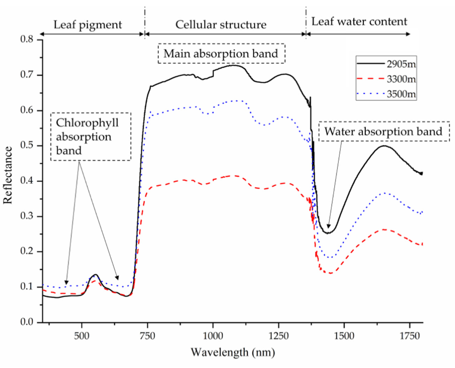
Рисунок 1. Спектры отражения листьев Quercus aquifolioides на разных высотах. Типичная кривая растительности имеет низкое отражение в видимом диапазоне, определяемое пигментами, «красный край» между видимым и NIR-диапазоном и высокое отражение в NIR, связанное с клеточной структурой. После 1300 нм характеристики преимущественно определяются содержанием воды. Перепечатано из [12]. ©2020 Zhu et al.
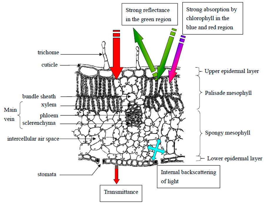
Рисунок 2. Схема поперечного среза листа и взаимодействия света с его анатомическими структурами. Пигменты, эпидермис, палисадная и губчатая паренхима, межклеточные пространства, трихомы, кутикула и устьица определяют поглощение, отражение, внутреннее рассеяние и пропускание света. Адаптировано из [23].
В NIR-области отражение главным образом определяется клеточной структурой и количеством межклеточных границ. Деградация тканей, потеря тургора и изменение водного содержания смещают спектральный отклик. Вода имеет выраженные полосы поглощения около 970, 1200, 1450, 1940 и 2500 нм; поэтому дефицит воды можно отслеживать по соответствующим диапазонам [23,24]. На уровне полога геометрия листьев, индекс листовой поверхности, почвенный фон и освещение усложняют сигнал [25,26], что требует стандартизации геометрии, эталонов отражения и условий съёмки.
Спектральные различия стресса нередко малы и перекрываются: разные заболевания, дефициты питания и водный стресс могут одинаково снижать хлорофилл или закрывать устьица. Поэтому отдельная длина волны редко обеспечивает достаточную специфичность. Эффективные системы объединяют множество полос, временную динамику, пространственную структуру изображения и алгоритмы классификации.
3. Датчики и сбор данных
Оптические методы охватывают электромагнитный спектр от UV до теплового IR (рисунок 3). Выбор датчика зависит от физического признака стресса, требуемого разрешения, скорости, стоимости и условий применения. Спектрометры измеряют интенсивность как функцию длины волны, тогда как камеры дополнительно сохраняют пространственное распределение. Проксимальные системы располагают рядом с листом или пологом, что повышает разрешение и снижает атмосферные помехи по сравнению с дистанционными платформами.
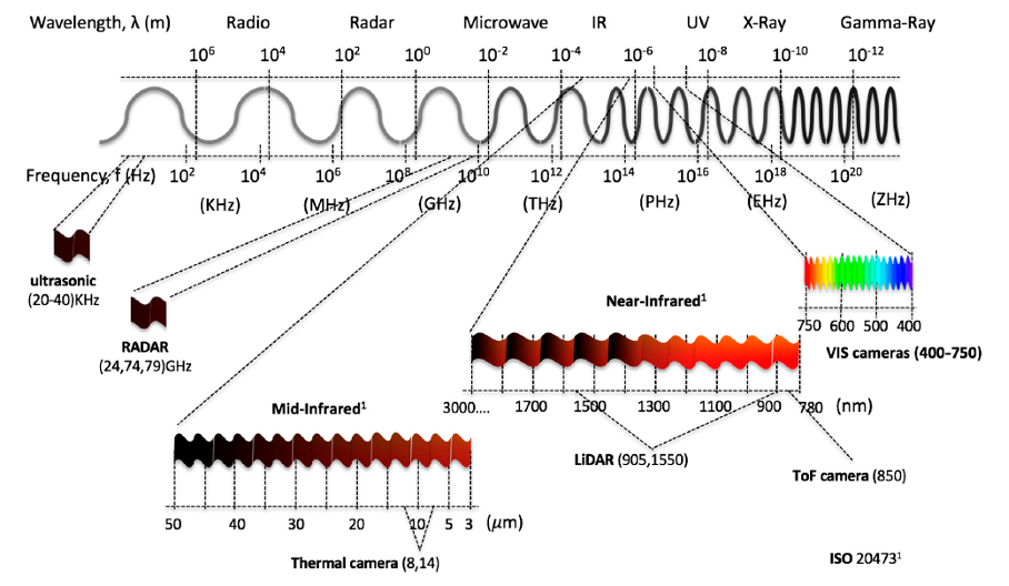
Рисунок 3. Диапазоны электромагнитного спектра и типичные области работы радиометров, радаров, микроволновых, инфракрасных, ультрафиолетовых и рентгеновских приборов, RGB-камер и тепловизоров [27].
3.1. Гиперспектральная визуализация
Гиперспектральная визуализация одновременно получает спектральную и пространственную информацию в десятках или сотнях смежных узких полос. Результат представляет собой гиперкуб с двумя пространственными и одним спектральным измерением. Он позволяет выявлять тонкие изменения пигментов, воды, структуры и метаболизма раньше видимых симптомов. Гиперспектральные методы применялись для обнаружения грибных, бактериальных и вирусных заболеваний, холода, засухи, засоления и дефицита питания [28–47].
Камеры работают по принципу точечного, построчного push-broom, спектрального сканирования либо snapshot-съёмки. Построчные системы дают высокое спектральное качество, но требуют относительного перемещения датчика и объекта и чувствительны к движению. Snapshot-камеры удобнее в поле, хотя обычно имеют меньше полос или меньшее пространственное разрешение. Современные миниатюрные приборы, например SpecimIQ (рисунок 4), объединяют камеру, освещение, сенсорный интерфейс и предварительную обработку.
Гиперспектральные данные требуют коррекции тёмного сигнала и белого эталона, удаления шумных полос, геометрической регистрации и нормализации. Высокая размерность увеличивает вычислительную нагрузку и риск переобучения, поэтому применяют выбор информативных длин волн, главные компоненты, частичные наименьшие квадраты и машинное обучение. Несмотря на стоимость, объём данных и необходимость контролировать освещение, гиперспектральная съёмка остаётся одним из наиболее чувствительных методов раннего выявления.
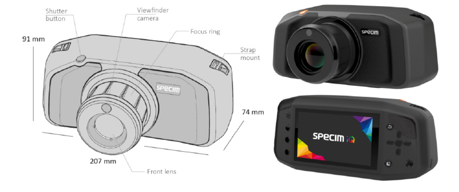
Рисунок 4. Миниатюрная гиперспектральная камера SpecimIQ (Specim Ltd., Оулу, Финляндия). Перепечатано из [52].
3.2. Мультиспектральная визуализация и спектроскопия
Мультиспектральные системы регистрируют ограниченное число широких или узких полос, выбранных по физиологической значимости. Они дешевле, быстрее и проще гиперспектральных и пригодны для портативной диагностики. Фильтры, светодиоды и недорогие фотодиоды позволяют создавать специальные приборы для конкретной культуры или стресса. Полосы видимого и NIR-диапазона использовались для пятнистости арахиса, серой гнили, болезней картофеля и пшеницы, водного и питательного стресса [53–60].
Смартфонные системы особенно перспективны: встроенная камера обеспечивает RGB-каналы, а внешнее освещение и фильтры расширяют диапазон. Портативный NIR-мониторинг смартфоном выявлял стресс растений [60], а устройство с держателем листа обеспечивало воспроизводимую геометрию при измерении риса (рисунок 5). Ограничениями остаются различия камер, автоматическая экспозиция и баланс белого, внешнее освещение и необходимость калибровки.
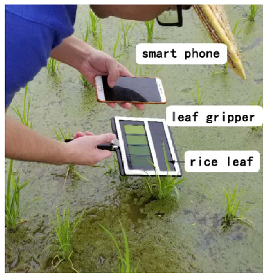
Рисунок 5. Смартфон, держатель и лист риса при полевом измерении оптических характеристик. Система фиксирует положение листа и расстояние до камеры для воспроизводимого анализа. Перепечатано из [60].
3.3. RGB-визуализация
RGB-камеры наиболее доступны и фиксируют признаки, видимые человеку: изменение цвета, хлороз, некроз, пятна, увядание и форму поражения. Высокое разрешение, низкая цена и наличие камер в смартфонах делают метод подходящим для массового мониторинга. Сегментация и анализ цвета применялись для болезней картофеля и пшеницы, оценки повреждения, дефицита азота и других стрессов [61–65].
Изображения RGB сильно зависят от освещения, теней, фона и модели камеры. Цветовые эталоны, контролируемые боксы, постоянная дистанция и перевод из RGB в HSV, Lab или нормализованные цветовые пространства уменьшают вариабельность. RGB обычно выявляет стресс позже спектральных или флуоресцентных методов, поскольку заметные симптомы формируются после физиологических изменений, однако автоматическое распознавание делает его ценным для дешёвой диагностики.
3.4. Тепловизионная съёмка и термография
Тепловизоры регистрируют излучение в длинноволновом IR и оценивают температуру поверхности. Закрытие устьиц снижает транспирационное охлаждение и повышает температуру листа; поэтому термография чувствительна к дефициту воды, солевому стрессу и заболеваниям, нарушающим устьичную регуляцию. Метод использовали для серой гнили винограда, засухи кукурузы, парши яблони и оценки проводимости устьиц [66–71].
На температуру влияют воздух, радиация, ветер, влажность, угол листа и фон, поэтому нужны эталоны влажного и сухого листа, нормализованные индексы и одновременно измеряемые метеоданные. Современные компактные тепловые модули подключаются к смартфону, но их пространственное разрешение ниже RGB. Совмещение тепловых и видимых изображений облегчает локализацию растений.
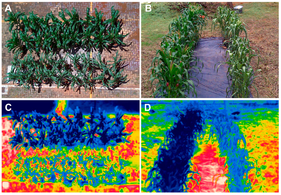
Рисунок 6. Термографические данные для оценки засухи у кукурузы: обычные изображения растений при разных режимах орошения и соответствующие карты температуры. Температурные различия отражают транспирацию и водный статус [68].
3.5. Флуоресцентная спектроскопия
После поглощения фотона молекула может испустить свет большей длины волны. У растений важнейшими источниками являются хлорофилл и фенольные соединения. Хлорофилл флуоресцирует преимущественно в красной и дальней красной областях около 690 и 740 нм; сине-зелёная флуоресценция связана, среди прочего, с клеточными стенками и фенолами [72–74]. Стресс изменяет фотохимию фотосистемы II, тушение энергии, содержание пигментов и тем самым интенсивность и кинетику флуоресценции.
Импульсно-амплитудно-модулированные флуорометры измеряют минимальную и максимальную флуоресценцию, переменную флуоресценцию и квантовый выход. Отношение Fv/Fm широко используется как показатель максимальной эффективности фотосистемы II [75,76]. Лазерно-индуцированная спектроскопия выявляла вирусные, бактериальные и грибные инфекции и гербицидный стресс [77–86]. Её преимущества — чувствительность и быстрота; ограничения — необходимость возбуждающего источника, контроль расстояния и ориентации и влияние темновой адаптации.
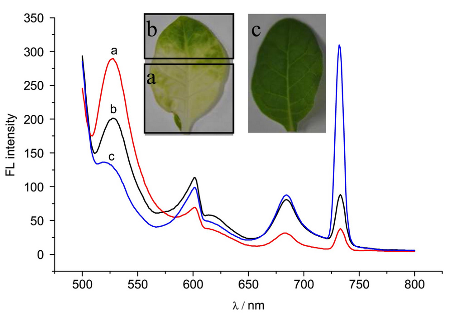
Рисунок 7. Спектры флуоресцентного излучения листьев при возбуждении 488 нм: (a) хлоротический участок листа табака; (b) здоровый участок того же листа; (c) здоровый лист. Различия пиков отражают изменение пигментов и фотосинтетического аппарата [72].
3.6. Флуоресцентная визуализация
Флуоресцентная визуализация формирует пространственные карты параметров по всему листу и выявляет неоднородные очаги до появления видимых симптомов. Многоцветные системы регистрируют сине-зелёную, красную и дальне-красную флуоресценцию; системы кинетики хлорофилла строят карты F0, Fm, Fv/Fm, эффективного квантового выхода и нефотохимического тушения [79–93]. Они применялись для засухи, дефицита азота, вирусов, грибов, бактерий и действия гербицидов.
Пример картирования растений при 0, 50 и 100% азотного питания показывает, что сочетания каналов 370F440, 370F740, 370F689, 447F689 и 447F740 визуально разделяют уровни питания (рисунок 8). Метод чувствителен, но требует стабильного возбуждения, фильтров, калибровки и управления большими массивами изображений.
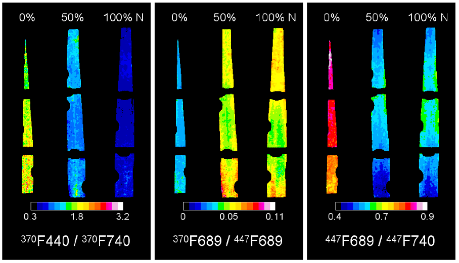
Рисунок 8. Флуоресцентные изображения растений при трёх уровнях азотного питания в различных комбинациях длин волн возбуждения и регистрации; цветовые карты показывают пространственные различия сигнала [84].
3.7. Слияние датчиков
Ни один оптический метод не является универсально специфичным. Объединение RGB, спектральной, тепловой и флуоресцентной информации повышает точность и позволяет различать стрессы с похожими признаками. Слияние возможно на уровне исходных данных, признаков или решений классификаторов [94–101]. Оно улучшает устойчивость к изменению освещения и стадиям развития, однако требует регистрации изображений, синхронизации, нормализации масштабов и более сложной вычислительной системы.
Таблица 1. Оптические методы выявления стресса растений.
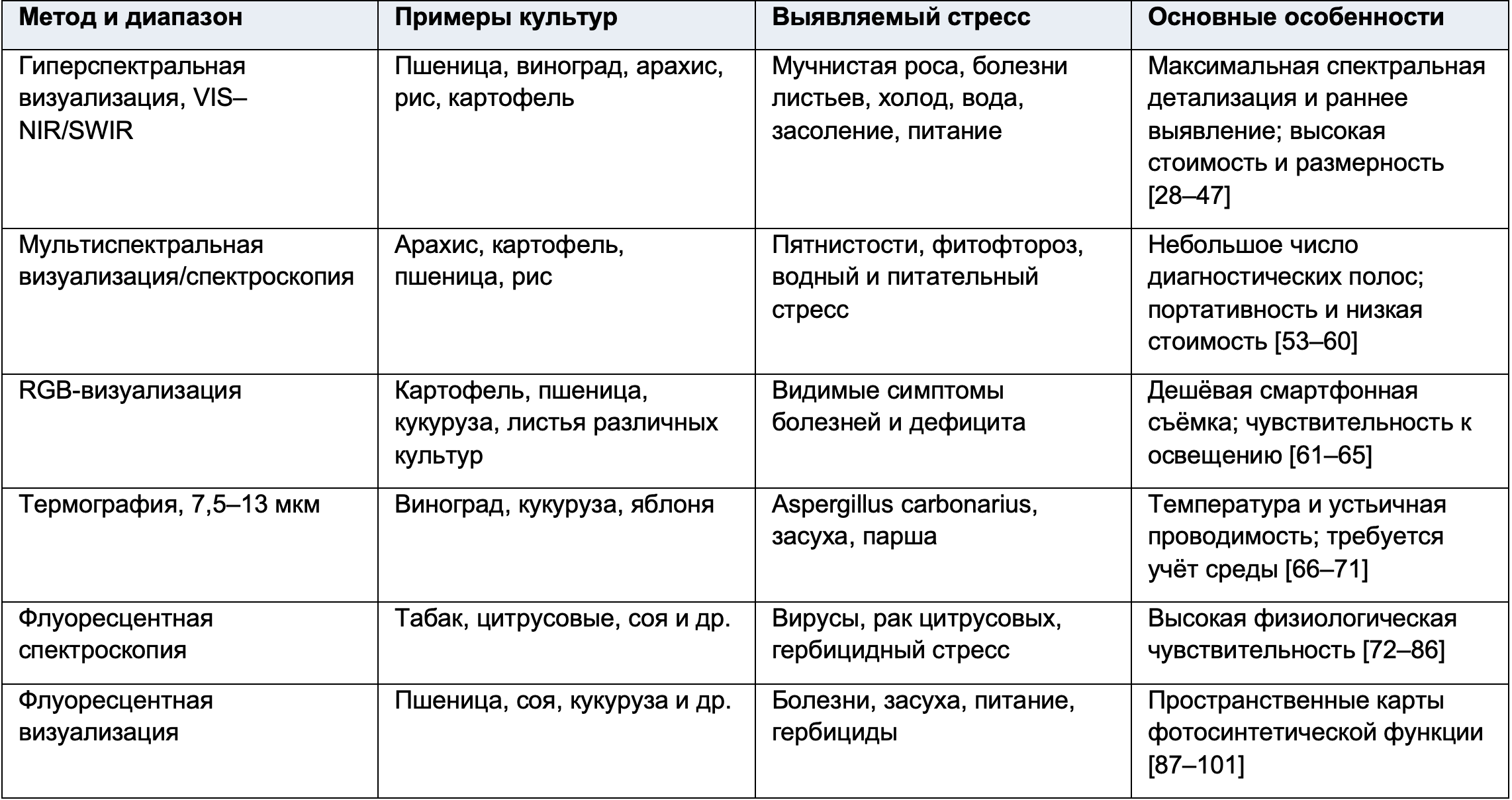
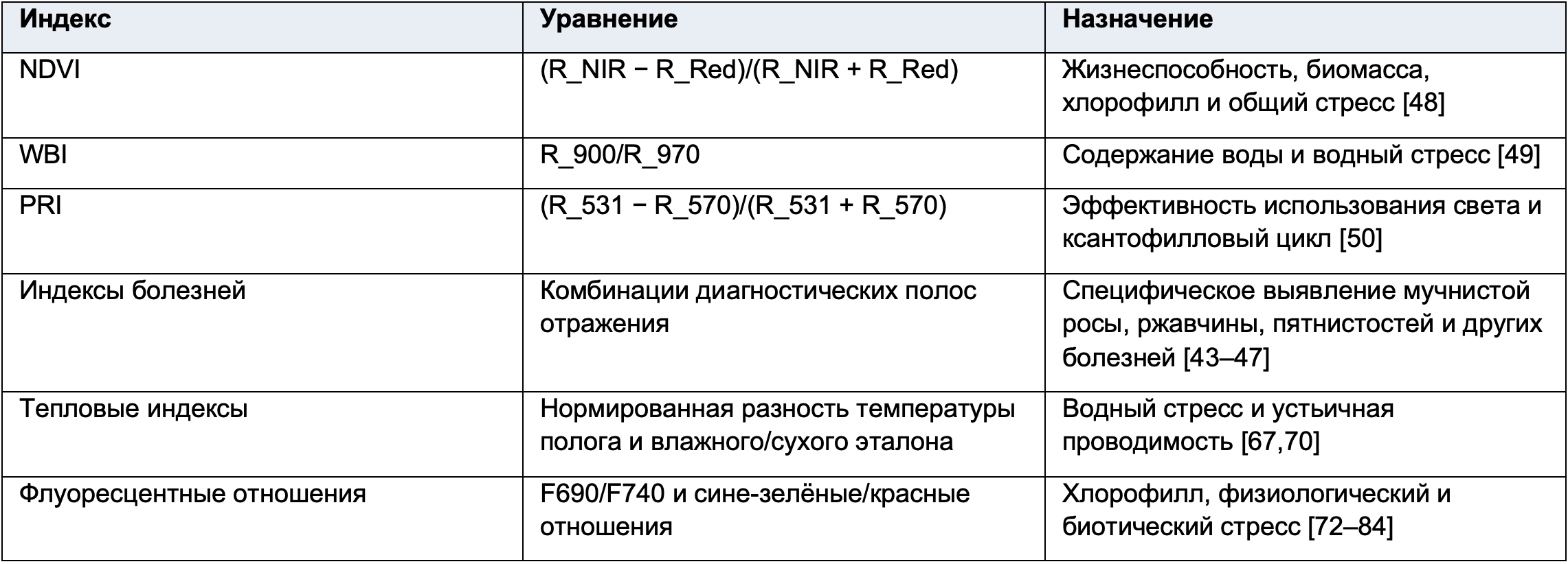
Таблица 2. Уравнения и применение вегетационных и болезненных индексов.
4. Машинное обучение для обработки данных
Оптические данные необходимо преобразовать в воспроизводимое решение о наличии и типе стресса. Типичный процесс включает сбор размеченных данных, предварительную обработку, извлечение признаков, обучение алгоритма, проверку на независимых образцах и классификацию новых наблюдений (рисунок 9). Качество модели определяется не только алгоритмом, но и репрезентативностью выборки, корректным разделением обучающих и тестовых данных и предотвращением утечки информации.
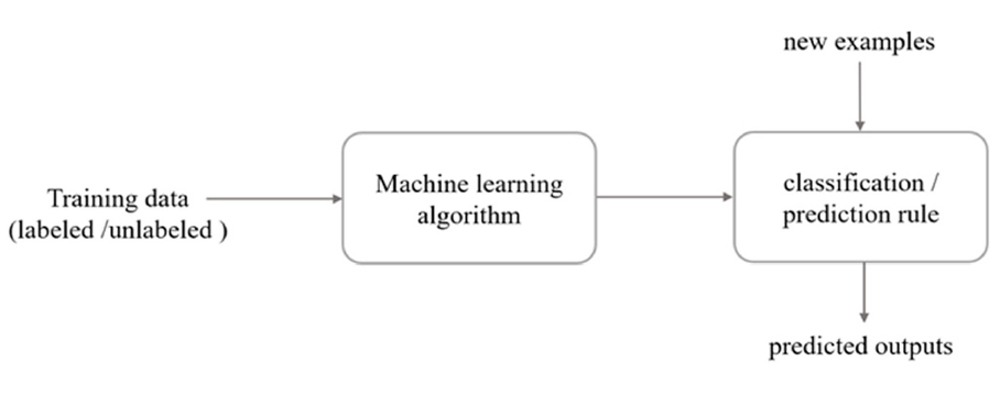
Рисунок 9. Упрощённая схема машинного обучения: размеченные или неразмеченные обучающие данные поступают в алгоритм, который формирует правило прогнозирования; новые примеры преобразуются в предсказанные классы или значения. Перепечатано из [104]. ©2018 Liakos et al.
4.1. Предварительная обработка
4.1.1. Преобразование цветового пространства
RGB удобно для отображения, но его каналы коррелированы и зависят от освещения. Преобразование в HSV/HSL отделяет оттенок и насыщенность от яркости; CIE Lab и Luv приближают цветовые расстояния к человеческому восприятию; YCbCr отделяет яркость от цветоразностных компонентов. Выбор пространства повышает различимость поражённой и здоровой ткани и облегчает сегментацию (рисунок 10).
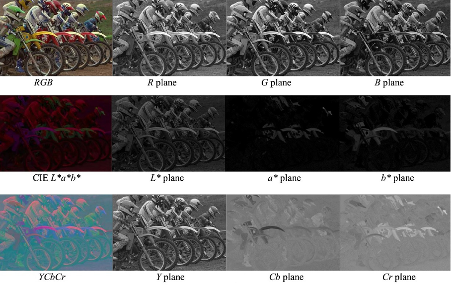
Рисунок 10. Представление RGB-изображения в других цветовых пространствах и по отдельным компонентам: R, G, B; H, S, V; L*, a*, b*; Y, Cb, Cr. Адаптировано из [61].
4.1.2. Снижение размерности
Гиперспектральные кубы содержат сотни сильно коррелированных переменных. Анализ главных компонент (PCA) преобразует их в ортогональные компоненты, сохраняющие большую часть дисперсии; частичные наименьшие квадраты ориентируют снижение на прогнозируемый отклик; линейный дискриминантный анализ максимизирует разделение классов. Выбор длин волн и методы отбора признаков уменьшают время, шум и переобучение и позволяют перейти к дешёвому мультиспектральному прибору.
4.1.3. Сегментация
Сегментация отделяет растение или поражённую область от почвы, фона и здоровой ткани. Используют пороговую обработку цвета и индексов, кластеризацию k-средних, выращивание областей, водораздел, активные контуры, морфологические операции и обучаемые классификаторы пикселей. Результат критически влияет на извлекаемые площадь, цвет, текстуру и температуру. На рисунке 11 показано выделение растений при различных условиях и методах.
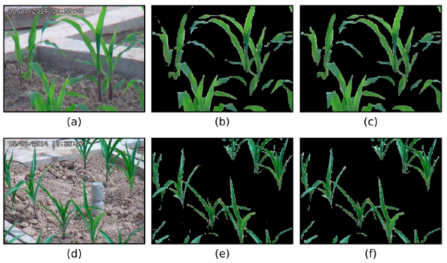
Рисунок 11. Визуализация сегментации растений: исходные полевые изображения и соответствующие бинарные/цветовые маски, отделяющие листья от почвы и фона [103].
4.1.4. Извлечение признаков
Признаки включают средние и распределения спектрального отражения, индексы, цветовые моменты, гистограммы, форму, площадь поражения, края и текстуру. Текстурные матрицы совместной встречаемости уровней серого дают контраст, корреляцию, энергию и однородность. Локальные бинарные шаблоны, вейвлеты и частотные преобразования фиксируют пятнистость и пространственный масштаб. Для временных серий используют скорость изменения. Глубокое обучение способно извлекать признаки автоматически, но требует большего объёма данных.
4.2. Алгоритмы машинного обучения для классификации
Задача может быть бинарной (здоровое/стресс), многоклассовой (тип или тяжесть стресса) либо регрессионной (содержание воды, азота, температура, процент поражения). Производительность оценивают точностью, чувствительностью, специфичностью, precision, recall, F1, ROC-AUC и ошибками регрессии. При несбалансированных классах одной общей точности недостаточно. Перекрёстная проверка должна разделять независимые растения, даты или поля, а не соседние пиксели одного изображения.
4.2.1. Метод опорных векторов (SVM)
SVM строит разделяющую гиперплоскость с максимальным зазором между классами. Ядерные функции, включая линейную, полиномиальную и радиально-базисную, позволяют нелинейное разделение (рисунок 12). SVM эффективен при малом числе образцов и большом числе спектральных признаков и широко применялся для болезней и абиотического стресса [29,33,35,36,43–47,94]. Требуются масштабирование, настройка штрафа и параметров ядра; вероятность и многоклассовость реализуются дополнительными схемами.
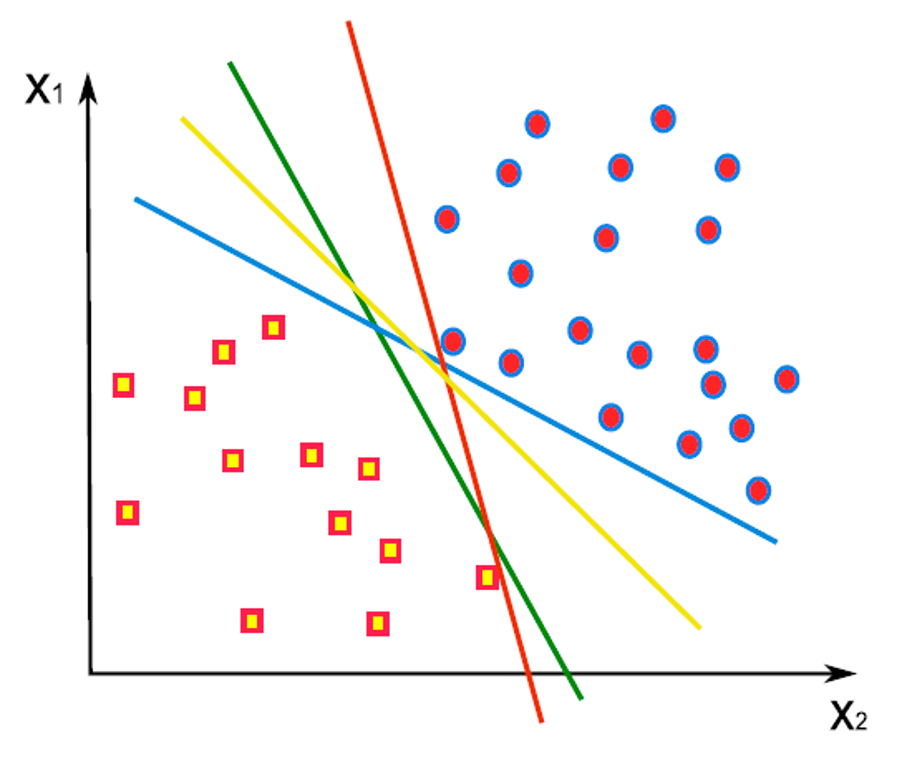
Рисунок 12. Границы решений SVM для двух классов в пространстве признаков X₁ и X₂. Разные разделяющие прямые дают различный зазор; оптимальная граница максимизирует расстояние до ближайших опорных векторов.
4.2.2. Искусственная нейронная сеть (ANN)
ANN состоит из взаимосвязанных узлов во входном, одном или нескольких скрытых и выходном слоях. Веса обучаются обратным распространением ошибки. Сети моделируют сложные нелинейные связи спектров, изображений и стресса и применялись для классификации болезней, водного и питательного статуса. Архитектуры включают многослойный персептрон, радиально-базисные сети, рекуррентные и самоорганизующиеся карты (рисунок 13). Результат зависит от объёма и разнообразия данных, регуляризации, нормализации и настройки; малые наборы повышают риск переобучения.
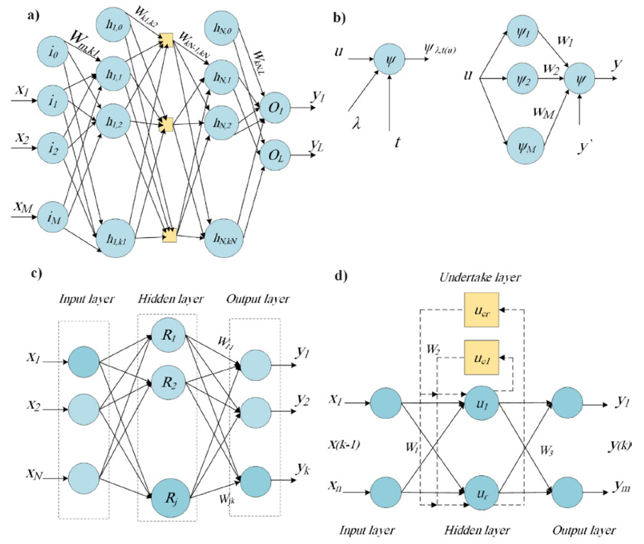
Рисунок 13. Структуры четырёх типов нейронных сетей: (a) многослойная полносвязная сеть; (b) простой нейрон и его взвешенное суммирование; (c) сеть с радиально-базисным скрытым слоем; (d) рекуррентная/динамическая структура. Схемы показывают входные, скрытые и выходные узлы и веса связей.
4.2.3. Глубокое обучение
Свёрточные нейронные сети (CNN) обучают пространственные фильтры непосредственно по изображениям и заменяют ручное извлечение признаков. Повторяющиеся свёртки, нелинейности и пулинг формируют иерархию от краёв и текстур до симптомов; полносвязный выход выполняет классификацию или регрессию. Перенос обучения из крупных наборов изображений уменьшает потребность в размеченных сельскохозяйственных данных. Трёхмерные CNN совместно обрабатывают пространственные и спектральные измерения гиперкубов.
Глубокие модели дают высокую точность, но требуют вычислительных ресурсов, большого разнообразия данных и интерпретируемости. Аугментация, dropout, ранняя остановка и внешняя валидация снижают переобучение. Для портативных систем важны облегчённые архитектуры, квантование и выполнение на смартфоне или периферийном устройстве.
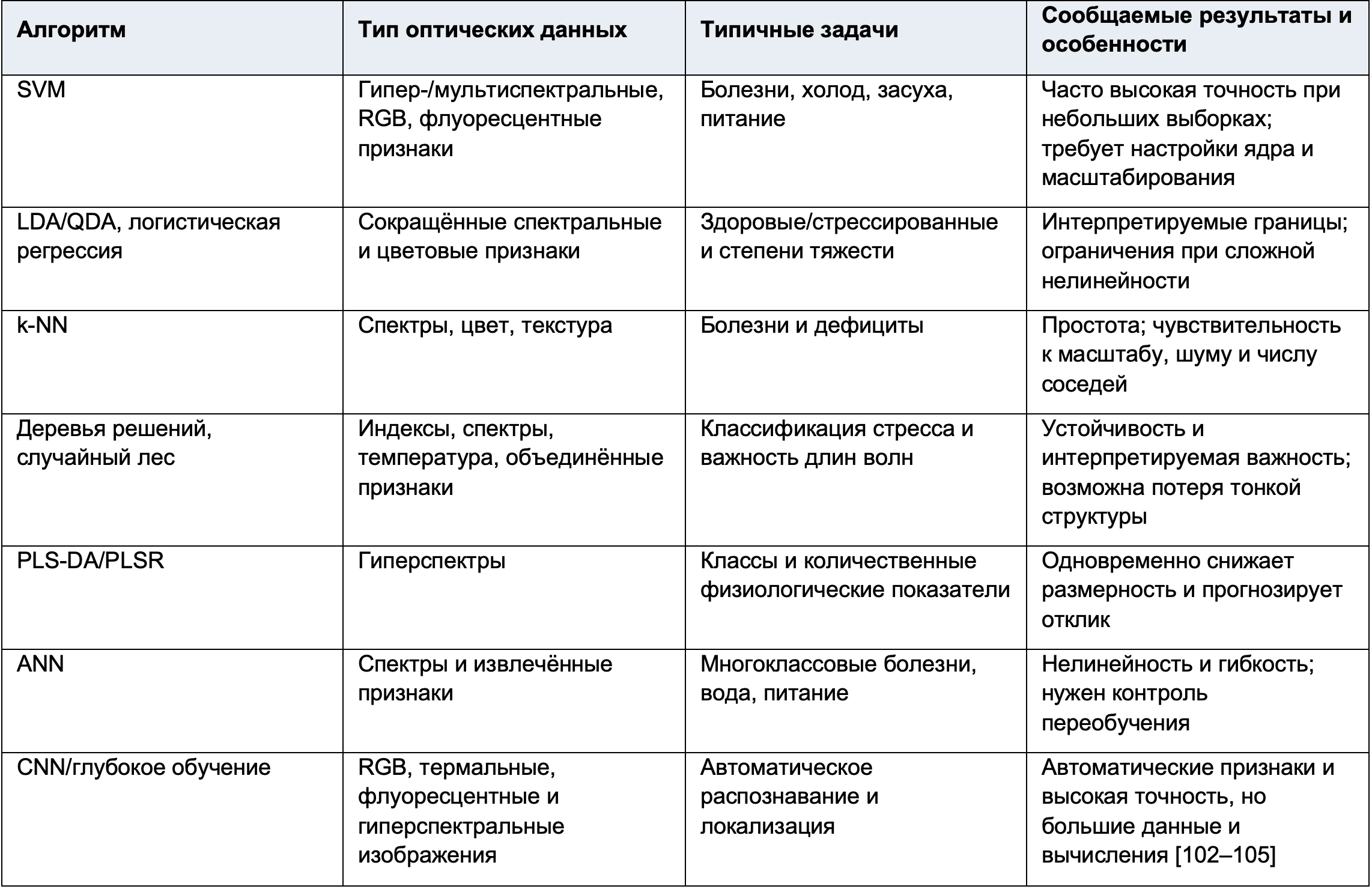
Таблица 3. Алгоритмы машинного обучения, применявшиеся для выявления стресса растений.
5. Заключительные замечания
Оптические датчики способны быстро и неразрушающе выявлять биотические и абиотические стрессы по изменениям пигментов, структуры, воды, температуры и фотосинтетической функции. Миниатюризация гиперспектральных камер, дешёвые мультиспектральные и тепловые модули и резкое улучшение смартфонов делают проксимальный мониторинг всё доступнее. Гиперспектральная съёмка обеспечивает наибольшую спектральную полноту; мультиспектральная — практический компромисс; RGB — минимальную стоимость; термография чувствительна к транспирации; флуоресценция непосредственно характеризует фотохимию. Слияние датчиков компенсирует ограничения отдельных методов.
Машинное обучение необходимо для преобразования сложных изображений и спектров в объективную идентификацию и классификацию. SVM, ANN, деревья, дискриминантные методы и глубокие CNN показали высокую точность, но опубликованные результаты нельзя считать автоматически переносимыми в практику. Небольшие и однородные наборы, случайное разделение пикселей одного растения, отсутствие независимой полевой проверки и различия оборудования способны завышать показатели.
Для практического применения нужны стандартизированные протоколы сбора и калибровки, открытые наборы данных с несколькими культурами, генотипами, стадиями и сезонами, а также проверка при естественном освещении и смешанных стрессах. Модели должны различать причины со сходными симптомами, оценивать неопределённость и объяснять решение. Важны продольные наблюдения, определяющие, насколько раньше видимых симптомов возникает надёжный сигнал и улучшает ли это управленческий результат.
Перспективные системы объединят смартфон или портативный датчик, контролируемое освещение, автоматическую проверку качества, встроенную модель и понятное агрономическое действие. Обработка на устройстве уменьшит задержку и зависимость от связи. Облачные обновления и перенос обучения позволят адаптировать модели к новым культурам и регионам. Для успешного внедрения устройство должно быть не только точным, но и дешёвым, быстрым, надёжным, простым, воспроизводимым и полезным в конкретной системе управления.
В целом сочетание портативных оптических датчиков и машинного обучения обладает значительным потенциалом для раннего выявления стресса, сокращения потерь, более точного применения воды, удобрений и средств защиты и повышения устойчивости сельского хозяйства. Междисциплинарное сотрудничество инженеров, специалистов по растениям, патологов, аналитиков данных и производителей необходимо для перехода от лабораторных демонстраций к надёжным полевым инструментам.
Вклад авторов: концепция — A.V.Z. и J.-Y.Y.; обзор литературы и сбор данных — A.V.Z.; подготовка первоначального текста — A.V.Z.; рецензирование и редактирование — A.V.Z. и J.-Y.Y.; визуализация — A.V.Z.; руководство и администрирование проекта — J.-Y.Y. Все авторы прочли опубликованную версию рукописи и согласились с ней.
Финансирование: исследование не получало внешнего финансирования.
Благодарности: авторы выражают благодарность за поддержку и материалы, указанные в исходной публикации.
Конфликт интересов: авторы заявляют об отсутствии конфликта интересов.
Литература
Библиографические записи сохранены в оригинальном написании для точной идентификации источников.
1. The Future of Food and Agriculture: Trends and Challenges; Food and Agriculture Organization of the United Nations: Rome, Italy, 2017; ISBN 978-92-5-109551-5.
2. Savary, S.; Bregaglio, S.; Willocquet, L.; Gustafson, D.; Mason D’Croz, D.; Sparks, A.; Castilla, N.; Djurle, A.; Allinne, C.; Sharma, M.; et al. Crop health and its global impacts on the components of food security. Food Secur. 2017, 9, 311–327. [CrossRef]
3. Savary, S.; Willocquet, L.; Pethybridge, S.J.; Esker, P.; McRoberts, N.; Nelson, A. The global burden of pathogens and pests on major food crops. Nat. Ecol. Evol. 2019, 3, 430–439. [CrossRef] [PubMed]
4. McDonald, B.A.; Stukenbrock, E.H. Rapid emergence of pathogens in agro-ecosystems: Global threats to agricultural sustainability and food security. Philos. Trans. R. Soc. B Biol. Sci. 2016, 371, 20160026. [CrossRef] [PubMed]
5. Shaw, M.W.; Osborne, T.M. Geographic distribution of plant pathogens in response to climate change: Pathogen distributions and climate. Plant Pathol. 2011, 60, 31–43. [CrossRef]
6. Yeo, A. Predicting the interaction between the effects of salinity and climate change on crop plants. Sci. Hortic. 1998, 78, 159–174. [CrossRef]
7. Sui, X.; Zheng, Y.; Li, R.; Padmanabhan, C.; Tian, T.; Groth-Helms, D.; Keinath, A.P.; Fei, Z.; Wu, Z.; Ling, K.-S. Molecular and Biological Characterization of Tomato mottle mosaic virus and Development of RT-PCR Detection. Plant Dis. 2017, 101, 704–711. [CrossRef]
8. Cimmino, A.; Iannaccone, M.; Petriccione, M.; Masi, M.; Evidente, M.; Capparelli, R.; Scortichini, M.; Evidente, A. An ELISA method to identify the phytotoxic Pseudomonas syringae pv. actinidiae exopolysaccharides: A tool for rapid immunochemical detection of kiwifruit bacterial canker. Phytochem. Lett. 2017, 19, 136–140. [CrossRef]
9. Andolfi, A.; Cimmino, A.; Evidente, A.; Iannaccone, M.; Capparelli, R.; Mugnai, L.; Surico, G. A New Flow Cytometry Technique to Identify Phaeomoniella chlamydospora Exopolysaccharides and Study Mechanisms of Esca Grapevine Foliar Symptoms. Plant Dis. 2009, 93, 680–684. [CrossRef]
10. McKenzie, D.B.; Hossner, L.R.; Newton, R.J. Sorghum cultivar evaluation for iron chlorosis resistance by visual scores. J. Plant Nutr. 1984, 7, 677–685. [CrossRef]
11. Naik, H.S.; Zhang, J.; Lofquist, A.; Assefa, T.; Sarkar, S.; Ackerman, D.; Singh, A.; Singh, A.K.; Ganapathysubramanian, B. A real-time phenotyping framework using machine learning for plant stress severity rating in soybean. Plant Methods 2017, 13, 23. [CrossRef]
12. Zhu, J.; He, W.; Yao, J.; Yu, Q.; Xu, C.; Huang, H.; Mhae, B.; Jandug, C. Spectral Reflectance Characteristics and Chlorophyll Content Estimation Model of Quercus aquifolioides Leaves at Different Altitudes in Sejila Mountain. Appl. Sci. 2020, 10, 3636. [CrossRef]
13. Lichtenthaler, H.K.; Gitelson, A.; Lang, M. Non-Destructive Determination of Chlorophyll Content of Leaves of a Green and an Aurea Mutant of Tobacco by Reflectance Measurements. J. Plant Physiol. 1996, 148, 483–493. [CrossRef]
14. Gitelson, A.A.; Zur, Y.; Chivkunova, O.B.; Merzlyak, M.N. Assessing Carotenoid Content in Plant Leaves with Reflectance Spectroscopy. Photochem. Photobiol. 2002, 75, 272–281. [CrossRef]
15. Vilfan, N.; Van der Tol, C.; Yang, P.; Wyber, R.; Malenovský, Z.; Robinson, S.A.; Verhoef, W. Extending Fluspect to simulate xanthophyll driven leaf reflectance dynamics. Remote Sens. Environ. 2018, 211, 345–356. [CrossRef]
16. Bone, R.A.; Lee, D.W.; Norman, J.M. Epidermal cells functioning as lenses in leaves of tropical rain-forest shade plants. Appl. Opt. 1985, 24, 1408. [CrossRef] [PubMed]
17. Grant, L.; Daughtry, C.S.T.; Vanderbilt, V.C. Polarized and specular reflectance variation with leaf surface features. Physiol. Plant. 1993, 88, 1–9. [CrossRef]
18. Ehleringer, J.; Bjorkman, O.; Mooney, H.A. Leaf Pubescence: Effects on Absorptance and Photosynthesis in a Desert Shrub. Science 1976, 192, 376–377. [CrossRef]
19. Bornman, J.F.; Vogelmann, T.C. Effect of UV-B Radiation on Leaf Optical Properties Measured with Fibre Optics. J. Exp. Bot. 1991, 42, 547–554. [CrossRef]
20. Peñuelas, J.; Filella, I.; Biel, C.; Serrano, L.; Savé, R. The reflectance at the 950–970 nm region as an indicator of plant water status. Int. J. Remote Sens. 1993, 14, 1887–1905. [CrossRef]
21. Liew, O.; Chong, P.; Li, B.; Asundi, A. Signature Optical Cues: Emerging Technologies for Monitoring Plant Health. Sensors 2008, 8, 3205–3239. [CrossRef]
22. Sawinski, K.; Mersmann, S.; Robatzek, S.; Böhmer, M. Guarding the Green: Pathways to Stomatal Immunity. Mol. Plant-Microbe Interact. 2013, 26, 626–632. [CrossRef] [PubMed]
23. Fourty, T.; Baret, F.; Jacquemoud, S.; Schmuck, G.; Verdebout, J. Leaf optical properties with explicit description of its biochemical composition: Direct and inverse problems. Remote Sens. Environ. 1996, 56, 104–117. [CrossRef]
24. de Lima, R.B.; dos Santos, T.B.; Vieira, L.G.E.; de Lourdes Lúcio Ferrarese, M.; Ferrarese-Filho, O.; Donatti, L.; Boeger, M.R.T.; de Oliveira Petkowicz, C.L. Salt stress alters the cell wall polysaccharides and anatomy of coffee (Coffea arabica L.) leaf cells. Carbohydr. Polym. 2014, 112, 686–694. [CrossRef] [PubMed]
25. Allen, W.A.; Richardson, A.J. Interaction of Light with a Plant Canopy. J. Opt. Soc. Am. 1968, 58, 1023. [CrossRef]
26. Carter, G.A. Responses of Leaf Spectral Reflectance to Plant Stress. Am. J. Bot. 1993, 80, 239–243. [CrossRef]
27. Rosique, F.; Navarro, P.J.; Fernández, C.; Padilla, A. A Systematic Review of Perception System and Simulators for Autonomous Vehicles Research. Sensors 2019, 19, 648. [CrossRef]
28. Pandey, P.; Ge, Y.; Stoerger, V.; Schnable, J.C. High Throughput In vivo Analysis of Plant Leaf Chemical Properties Using Hyperspectral Imaging. Front. Plant Sci. 2017, 8, 1348. [CrossRef]
29. Rumpf, T.; Mahlein, A.-K.; Steiner, U.; Oerke, E.-C.; Dehne, H.-W.; Plümer, L. Early detection and classification of plant diseases with Support Vector Machines based on hyperspectral reflectance. Comput. Electron. Agric. 2010, 74, 91–99. [CrossRef]
30. Yang, W.; Yang, C.; Hao, Z.; Xie, C.; Li, M. Diagnosis of Plant Cold Damage Based on Hyperspectral Imaging and Convolutional Neural Network. IEEE Access 2019, 7, 118239–118248. [CrossRef]
31. Zovko, M.; Žibrat, U.; Knapič, M.; Kovačić, M.B.; Romić, D. Hyperspectral remote sensing of grapevine drought stress. Precis. Agric. 2019, 20, 335–347. [CrossRef]
32. Asaari, M.S.M.; Mertens, S.; Dhondt, S.; Inzé, D.; Wuyts, N.; Scheunders, P. Analysis of hyperspectral images for detection of drought stress and recovery in maize plants in a high-throughput phenotyping platform. Comput. Electron. Agric. 2019, 162, 749–758. [CrossRef]
33. Behmann, J.; Steinrücken, J.; Plümer, L. Detection of early plant stress responses in hyperspectral images. ISPRS J. Photogramm. Remote Sens. 2014, 93, 98–111. [CrossRef]
34. Zhang, J.; Yuan, L.; Pu, R.; Loraamm, R.W.; Yang, G.; Wang, J. Comparison between wavelet spectral features and conventional spectral features in detecting yellow rust for winter wheat. Comput. Electron. Agric. 2014, 100, 79–87. [CrossRef]
35. Cao, X.; Luo, Y.; Zhou, Y.; Duan, X.; Cheng, D. Detection of powdery mildew in two winter wheat cultivars using canopy hyperspectral reflectance. Crop Prot. 2013, 45, 124–131. [CrossRef]
36. Feng, X.; Zhan, Y.; Wang, Q.; Yang, X.; Yu, C.; Wang, H.; Tang, Z.; Jiang, D.; Peng, C.; He, Y. Hyperspectral imaging combined with machine learning as a tool to obtain high-throughput plant salt-stress phenotyping. Plant J. 2020, 101, 1448–1461. [CrossRef] [PubMed]
37. Ochoa, D.; Cevallos, J.; Vargas, G.; Criollo, R.; Romero, D.; Castro, R.; Bayona, O. Hyperspectral Imaging System for Disease Scanning on Banana Plants. In Proceedings of the Sensing for Agriculture and Food Quality and Safety VIII, Baltimore, MD, USA, 20–21 April 2016. [CrossRef]
38. Lowe, A.; Harrison, N.; French, A.P. Hyperspectral image analysis techniques for the detection and classification of the early onset of plant disease and stress. Plant Methods 2017, 13, 80. [CrossRef]
39. Brugger, A.; Behmann, J.; Paulus, S.; Luigs, H.-G.; Kuska, M.T.; Schramowski, P.; Kersting, K.; Steiner, U.; Mahlein, A.-K. Extending Hyperspectral Imaging for Plant Phenotyping to the UV-Range. Remote Sens. 2019, 11, 1401. [CrossRef]
40. Ryu, J.-H.; Jeong, H.; Cho, J. Performances of Vegetation Indices on Paddy Rice at Elevated Air Temperature, Heat Stress, and Herbicide Damage. Remote Sens. 2020, 12, 2654. [CrossRef]
41. Liu, Q.; Zhang, F.; Chen, J.; Li, Y. Water stress altered photosynthesis-vegetation index relationships for winter wheat. Agron. J. 2020, 112, 2944–2955. [CrossRef]
42. Ihuoma, S.O.; Madramootoo, C.A. Sensitivity of spectral vegetation indices for monitoring water stress in tomato plants. Comput. Electron. Agric. 2019, 163, 104860. [CrossRef]
43. Meng, R.; Lv, Z.; Yan, J.; Chen, G.; Zhao, F.; Zeng, L.; Xu, B. Development of Spectral Disease Indices for Southern Corn Rust Detection and Severity Classification. Remote Sens. 2020, 12, 3233. [CrossRef]
44. Huang, W.; Guan, Q.; Luo, J.; Zhang, J.; Zhao, J.; Liang, D.; Huang, L.; Zhang, D. New Optimized Spectral Indices for Identifying and Monitoring Winter Wheat Diseases. IEEE J. Sel. Top. Appl. Earth Obs. Remote Sens. 2014, 7, 2516–2524. [CrossRef]
45. Mahlein, A.-K.; Rumpf, T.; Welke, P.; Dehne, H.-W.; Plümer, L.; Steiner, U.; Oerke, E.-C. Development of spectral indices for detecting and identifying plant diseases. Remote Sens. Environ. 2013, 128, 21–30. [CrossRef]
46. Ashourloo, D.; Mobasheri, M.; Huete, A. Developing Two Spectral Disease Indices for Detection of Wheat Leaf Rust (Pucciniatriticina). Remote Sens. 2014, 6, 4723–4740. [CrossRef]
47. Heim, R.H.J.; Wright, I.J.; Allen, A.P.; Geedicke, I.; Oldeland, J. Developing a spectral disease index for myrtle rust (Austropuccinia psidii). Plant Pathol. 2019, 68, 738–745. [CrossRef]
48. Rouse, J.W.; Haas, R.H.; Schell, J.A.; Deering, D.W. Monitoring Vegetation Systems in the Great Plains with ERTS. NASA Goddard Space Flight Cent. 3d ERTS-1 Symp. 1974, 1, 9.
49. Penuelas, J.; Pinol, J.; Ogaya, R.; Filella, I. Estimation of plant water concentration by the reflectance Water Index WI (R900/R970). Int. J. Remote Sens. 1997, 18, 2869–2875. [CrossRef]
50. Gamon, J.A.; Peñuelas, J.; Field, C.B. A narrow-waveband spectral index that tracks diurnal changes in photosynthetic efficiency. Remote Sens. Environ. 1992, 41, 35–44. [CrossRef]
51. Balasundram, S.K.; Golhani, K.; Shamshiri, R.R.; Vadamalai, G. Precision Agriculture Technologies for Management of Plant Diseases. In Plant Disease Management Strategies for Sustainable Agriculture through Traditional and Modern Approaches; Ul Haq, I., Ijaz, S., Eds.; Sustainability in Plant and Crop Protection; Springer: Cham, Switzerland, 2020; Volume 13, pp. 259–278, ISBN 978-3-030-35954-6.
52. Behmann, J.; Acebron, K.; Emin, D.; Bennertz, S.; Matsubara, S.; Thomas, S.; Bohnenkamp, D.; Kuska, M.; Jussila, J.; Salo, H.; et al. Specim IQ: Evaluation of a New, Miniaturized Handheld Hyperspectral Camera and Its Application for Plant Phenotyping and Disease Detection. Sensors 2018, 18, 441. [CrossRef]
53. Chen, T.; Zhang, J.; Chen, Y.; Wan, S.; Zhang, L. Detection of peanut leaf spots disease using canopy hyperspectral reflectance. Comput. Electron. Agric. 2019, 156, 677–683. [CrossRef]
54. Veys, C.; Chatziavgerinos, F.; AlSuwaidi, A.; Hibbert, J.; Hansen, M.; Bernotas, G.; Smith, M.; Yin, H.; Rolfe, S.; Grieve, B. Multispectral imaging for presymptomatic analysis of light leaf spot in oilseed rape. Plant Methods 2019, 15, 4. [CrossRef]
55. Fahrentrapp, J. Detection of Gray Mold Leaf Infections Prior to Visual Symptom Appearance Using a Five-Band Multispectral Sensor. Front. Plant Sci. 2019, 10, 628. [CrossRef] [PubMed]
56. Cardim Ferreira Lima, M.; Krus, A.; Valero, C.; Barrientos, A.; del Cerro, J.; Roldán-Gómez, J.J. Monitoring Plant Status and Fertilization Strategy through Multispectral Images. Sensors 2020, 20, 435. [CrossRef] [PubMed]
57. Kitić, G.; Tagarakis, A.; Cselyuszka, N.; Panić, M.; Birgermajer, S.; Sakulski, D.; Matović, J. A new low-cost portable multispectral optical device for precise plant status assessment. Comput. Electron. Agric. 2019, 162, 300–308. [CrossRef]
58. Veys, C.; Hibbert, J.; Davis, P.; Grieve, B. An ultra-low-cost active multispectral crop diagnostics device. In Proceedings of the 2017 IEEE Sensors, Glasgow, UK, 29 October–1 November 2017; pp. 1–3.
59. Habibullah, M.; Mohebian, M.R.; Soolanayakanahally, R.; Bahar, A.N.; Vail, S.; Wahid, K.A.; Dinh, A. Low-Cost Multispectral Sensor Array for Determining Leaf Nitrogen Status. Nitrogen 2020, 1, 67–80. [CrossRef]
60. Chung, S.; Breshears, L.E.; Yoon, J.-Y. Smartphone near infrared monitoring of plant stress. Comput. Electron. Agric. 2018, 154, 93–98. [CrossRef]
61. Watchareeruetai, U.; Noinongyao, P.; Wattanapaiboonsuk, C.; Khantiviriya, P.; Duangsrisai, S. Identification of Plant Nutrient Deficiencies Using Convolutional Neural Networks; IEEE: Krabi, Thailand, 2018; pp. 1–4.
62. Islam, M.; Anh, D.; Wahid, K.; Bhowmik, P. Detection of potato diseases using image segmentation and multiclass support vector machine. In Proceedings of the 2017 IEEE 30th Canadian Conference on Electrical and Computer Engineering (CCECE), Windsor, ON, Canada, 30 April–3 May 2017; pp. 1–4.
63. Xie, X.; Zhang, X.; He, B.; Liang, D.; Zhang, D.; Huang, L. A System for Diagnosis of Wheat Leaf Diseases Based on Android Smartphone. In Proceedings of the International Symposium on Optical Measurement Technology and Instrumentation, Beijing, China, 9–11 May 2016. [CrossRef]
64. Mattupalli, C.; Moffet, C.; Shah, K.; Young, C. Supervised Classification of RGB Aerial Imagery to Evaluate the Impact of a Root Rot Disease. Remote Sens. 2018, 10, 917. [CrossRef]
65. Tao, M.; Ma, X.; Huang, X.; Liu, C.; Deng, R.; Liang, K.; Qi, L. Smartphone-based detection of leaf color levels in rice plants. Comput. Electron. Agric. 2020, 173, 105431. [CrossRef]
66. Mastrodimos, N.; Lentzou, D.; Templalexis, C.; Tsitsigiannis, D.I.; Xanthopoulos, G. Development of thermography methodology for early diagnosis of fungal infection in table grapes: The case of Aspergillus carbonarius. Comput. Electron. Agric. 2019, 165, 104972. [CrossRef]
67. Jones, H.G. Use of infrared thermometry for estimation of stomatal conductance as a possible aid to irrigation scheduling. Agric. For. Meteorol. 1999, 95, 139–149. [CrossRef]
68. Casari, R.; Paiva, D.; Silva, V.; Ferreira, T.; Souza, J.M.; Oliveira, N.; Kobayashi, A.; Molinari, H.; Santos, T.; Gomide, R.; et al. Using Thermography to Confirm Genotypic Variation for Drought Response in Maize. Int. J. Mol. Sci. 2019, 20, 2273. [CrossRef] [PubMed]
69. Oerke, E.-C.; Fröhling, P.; Steiner, U. Thermographic assessment of scab disease on apple leaves. Precis. Agric. 2011, 12, 699–715. [CrossRef]
70. Khorsandi, A.; Hemmat, A.; Mireei, S.A.; Amirfattahi, R.; Ehsanzadeh, P. Plant temperature-based indices using infrared thermography for detecting water status in sesame under greenhouse conditions. Agric. Water Manag. 2018, 204, 222–233. [CrossRef]
71. Petrie, P.R.; Wang, Y.; Liu, S.; Lam, S.; Whitty, M.A.; Skewes, M.A. The accuracy and utility of a low cost thermal camera and smartphone-based system to assess grapevine water status. Biosyst. Eng. 2019, 179, 126–139. [CrossRef]
72. Lang, M.; Siffel, P.; Braunová, Z.; Lichtenthaler, H.K. Investigations of the Blue-green Fluorescence Emission of Plant Leaves. Bot. Acta 1992, 105, 435–440. [CrossRef]
73. Krause, G.H.; Weis, E. Chlorophyll fluorescence as a tool in plant physiology: II. Interpretation of fluorescence signals. Photosynth. Res. 1984, 5, 139–157. [CrossRef]
74. Swarbrick, P.J.; Schulze-Lefert, P.; Scholes, J.D. Metabolic consequences of susceptibility and resistance (race-specific and broad-spectrum) in barley leaves challenged with powdery mildew. Plant Cell Environ. 2006, 29, 1061–1076. [CrossRef]
75. Lawson, T.; Vialet-Chabrand, S. Chlorophyll Fluorescence Imaging. In Photosynthesis; Covshoff, S., Ed.; Methods in Molecular Biology; Springer: New York, NY, USA, 2018; Volume 1770, pp. 121–140. ISBN 978-1-4939-7785-7.
76. Brooks, M.D.; Niyogi, K.K. Use of a Pulse-Amplitude Modulated Chlorophyll Fluorometer to Study the Efficiency of Photosynthesis in Arabidopsis Plants. In Chloroplast Research in Arabidopsis; Jarvis, R.P., Ed.; Methods in Molecular Biology; Humana Press: Totowa, NJ, USA, 2011; Volume 775, pp. 299–310, ISBN 978-1-61779-236-6.
77. Lei, R.; Du, Z.; Qiu, Y.; Zhu, S. The detection of hydrogen peroxide involved in plant virus infection by fluorescence spectroscopy: Detection of hydrogen peroxide in plant by fluorescence spectroscopy. Luminescence 2016, 31, 1158–1165. [CrossRef]
78. Lichtenthaler, H.K.; Rinderle, U. The Role of Chlorophyll Fluorescence in the Detection of Stress Conditions in Plants. CRC Crit. Rev. Anal. Chem. 1988, 19, S29–S85. [CrossRef]
79. Murchie, E.H.; Lawson, T. Chlorophyll fluorescence analysis: A guide to good practice and understanding some new applications. J. Exp. Bot. 2013, 64, 3983–3998. [CrossRef]
80. Gomes, M.T.G.; da Luz, A.C.; dos Santos, M.R.; do Carmo Pimentel Batitucci, M.; Silva, D.M.; Falqueto, A.R. Drought tolerance of passion fruit plants assessed by the OJIP chlorophyll a fluorescence transient. Sci. Hortic. 2012, 142, 49–56. [CrossRef]
81. Kalaji, H.M.; Oukarroum, A.; Alexandrov, V.; Kouzmanova, M.; Brestic, M.; Zivcak, M.; Samborska, I.A.; Cetner, M.D.; Allakhverdiev, S.I.; Goltsev, V. Identification of nutrient deficiency in maize and tomato plants by in vivo chlorophyll a fluorescence measurements. Plant Physiol. Biochem. 2014, 81, 16–25. [CrossRef] [PubMed]
82. Kalaji, H.M.; Baba,˛ W.; Gediga, K.; Goltsev, V.; Samborska, I.A.; Cetner, M.D.; Dimitrova, S.; Piszcz, U.; Bielecki, K.; Karmowska, K.; et al. Chlorophyll fluorescence as a tool for nutrient status identification in rapeseed plants. Photosynth. Res. 2018, 136, 329–343. [CrossRef] [PubMed]
83. Buschmann, C.; Lichtenthaler, H.K. Principles and characteristics of multi-colour fluorescence imaging of plants. J. Plant Physiol. 1998, 152, 297–314. [CrossRef]
84. Bürling, K.; Hunsche, M.; Noga, G. Use of blue–green and chlorophyll fluorescence measurements for differentiation between nitrogen deficiency and pathogen infection in winter wheat. J. Plant Physiol. 2011, 168, 1641–1648. [CrossRef]
85. Saleem, M.; Atta, B.M.; Ali, Z.; Bilal, M. Laser-induced fluorescence spectroscopy for early disease detection in grapefruit plants. Photochem. Photobiol. Sci. 2020, 19, 713–721. [CrossRef]
86. Lins, E.C.; Belasque, J.; Marcassa, L.G. Detection of citrus canker in citrus plants using laser induced fluorescence spectroscopy. Precis. Agric. 2009, 10, 319–330. [CrossRef]
87. Su, W.H.; Fennimore, S.A.; Slaughter, D.C. Fluorescence imaging for rapid monitoring of translocation behaviour of systemic markers in snap beans for automated crop/weed discrimination. Biosyst. Eng. 2019, 186, 156–167. [CrossRef]
88. Li, H.; Wang, P.; Weber, J.; Gerhards, R. Early Identification of Herbicide Stress in Soybean (Glycine max (L.) Merr.) Using Chlorophyll Fluorescence Imaging Technology. Sensors 2017, 18, 21. [CrossRef]
89. Dong, Z.; Men, Y.; Li, Z.; Zou, Q.; Ji, J. Chlorophyll fluorescence imaging as a tool for analyzing the effects of chilling injury on tomato seedlings. Sci. Hortic. 2019, 246, 490–497. [CrossRef]
90. Konanz, S.; Kocsányi, L.; Buschmann, C. Advanced Multi-Color Fluorescence Imaging System for Detection of Biotic and Abiotic Stresses in Leaves. Agriculture 2014, 4, 79–95. [CrossRef]
91. Chung, S.; Breshears, L.E.; Perea, S.; Morrison, C.M.; Betancourt, W.Q.; Reynolds, K.A.; Yoon, J.-Y. Smartphone-Based Paper Microfluidic Particulometry of Norovirus from Environmental Water Samples at the Single Copy Level. ACS Omega 2019, 4, 11180–11188. [CrossRef] [PubMed]
92. Takayama, K.; Nishina, H. Chlorophyll fluorescence imaging of the chlorophyll fluorescence induction phenomenon for plant health monitoring. Environ. Control Biol. 2009, 47, 101–109. [CrossRef]
93. Pérez-Bueno, M.L.; Pineda, M.; Barón, M. Phenotyping Plant Responses to Biotic Stress by Chlorophyll Fluorescence Imaging. Front. Plant Sci. 2019, 10, 1135. [CrossRef] [PubMed]
94. Moshou, D.; Bravo, C.; Oberti, R.; West, J.S.; Ramon, H.; Vougioukas, S.; Bochtis, D. Intelligent multi-sensor system for the detection and treatment of fungal diseases in arable crops. Biosyst. Eng. 2011, 108, 311–321. [CrossRef]
95. Berdugo, C.A.; Zito, R.; Paulus, S.; Mahlein, A.-K. Fusion of sensor data for the detection and differentiation of plant diseases in cucumber. Plant Pathol. 2014, 63, 1344–1356. [CrossRef]
96. Adhikari, R.; Li, C.; Kalbaugh, K.; Nemali, K. A low-cost smartphone controlled sensor based on image analysis for estimating whole-plant tissue nitrogen (N) content in floriculture crops. Comput. Electron. Agric. 2020, 169, 105173. [CrossRef]
97. Bebronne, R.; Carlier, A.; Meurs, R.; Leemans, V.; Vermeulen, P.; Dumont, B.; Mercatoris, B. In-field proximal sensing of septoria tritici blotch, stripe rust and brown rust in winter wheat by means of reflectance and textural features from multispectral imagery. Biosyst. Eng. 2020, 197, 257–269. [CrossRef]
98. Brambilla, M. Application of a low-cost RGB sensor to detect basil (Ocimum basilicum L.) nutritional status at pilot scale level. Precis. Agric. 2020, 20. [CrossRef]
99. Banerjee, K.; Krishnan, P.; Das, B. Thermal imaging and multivariate techniques for characterizing and screening wheat genotypes under water stress condition. Ecol. Indic. 2020, 119, 106829. [CrossRef]
100. Cen, H.; Weng, H.; Yao, J.; He, M.; Lv, J.; Hua, S.; Li, H.; He, Y. Chlorophyll Fluorescence Imaging Uncovers Photosynthetic Fingerprint of Citrus Huanglongbing. Front. Plant Sci. 2017, 8, 1509. [CrossRef] [PubMed]
101. Raji, S.N.; Subhash, N.; Ravi, V.; Saravanan, R.; Mohanan, C.N.; Nita, S.; Kumar, T.M. Detection of mosaic virus disease in cassava plants by sunlight-induced fluorescence imaging: A pilot study for proximal sensing. Int. J. Remote Sens. 2015, 36, 2880–2897. [CrossRef]
102. Singh, A.; Ganapathysubramanian, B.; Singh, A.K.; Sarkar, S. Machine Learning for High-Throughput Stress Phenotyping in Plants. Trends Plant Sci. 2016, 21, 110–124. [CrossRef] [PubMed]
103. Ramos-Giraldo, P.; Reberg-Horton, C.; Locke, A.M.; Mirsky, S.; Lobaton, E. Drought Stress Detection Using Low-Cost Computer Vision Systems and Machine Learning Techniques. IT Prof. 2020, 22, 27–29. [CrossRef]
104. Liakos, K.; Busato, P.; Moshou, D.; Pearson, S.; Bochtis, D. Machine Learning in Agriculture: A Review. Sensors 2018, 18, 2674. [CrossRef] [PubMed]
105. Tsaftaris, S.A.; Minervini, M.; Scharr, H. Machine Learning for Plant Phenotyping Needs Image Processing. Trends Plant Sci. 2016, 21, 989–991. [CrossRef] [PubMed]
106. Dhakate, M.; Ingole, A.B. Diagnosis of pomegranate plant diseases using neural network. In Proceedings of the 2015 Fifth National Conference on Computer Vision, Pattern Recognition, Image Processing and Graphics (NCVPRIPG), Patna, India, 16–19 December 2015; pp. 1–4.
107. Al Bashish, D.; Braik, M.; Bani-Ahmad, S. A framework for detection and classification of plant leaf and stem diseases. In Proceedings of the 2010 International Conference on Signal and Image Processing, Chennai, India, 15–17 December 2010; pp. 113–118.
108. Shrivastava, S.; Singh, S.K.; Hooda, D.S. Color sensing and image processing-based automatic soybean plant foliar disease severity detection and estimation. Multimed. Tools Appl. 2015, 74, 11467–11484. [CrossRef]
109. Kahu, S.Y.; Raut, R.B.; Bhurchandi, K.M. Review and evaluation of color spaces for image/video compression. Color Res. Appl. 2018, 44, 8–33. [CrossRef]
110. Lever, J.; Krzywinski, M.; Altman, N. Principal component analysis. Nat. Methods 2017, 14, 641–642. [CrossRef]
111. Lu, Y.; Yi, S.; Zeng, N.; Liu, Y.; Zhang, Y. Identification of rice diseases using deep convolutional neural networks. Neurocomputing 2017, 267, 378–384. [CrossRef]
112. Wold, S.; Esbensen, K.; Geladi, P. Principal Component Analysis. Chemom. Intell. Lab. Syst. 1987, 2, 37–52. [CrossRef]
113. Singh, V.; Misra, A.K. Detection of plant leaf diseases using image segmentation and soft computing techniques. Inf. Process. Agric. 2016, 4, 41–49. [CrossRef]
114. Ma, J.; Du, K.; Zheng, F.; Zhang, L.; Gong, Z.; Sun, Z. A recognition method for cucumber diseases using leaf symptom images based on deep convolutional neural network. Comput. Electron. Agric. 2018, 154, 18–24. [CrossRef]
115. Zhuang, S.; Wang, P.; Jiang, B.; Li, M.; Gong, Z. Early detection of water stress in maize based on digital images. Comput. Electron. Agric. 2017, 140, 461–468. [CrossRef]
116. Yue, J.; Li, Z.; Liu, L.; Fu, Z. Content-based image retrieval using color and texture fused features. Math. Comput. Model. 2011, 54, 1121–1127. [CrossRef]
117. Ojala, T.; Pietikainen, M.; Maenpaa, T. Multiresolution gray-scale and rotation invariant texture classification with local binary patterns. IEEE Trans. Pattern Anal. Mach. Intell. 2002, 24, 971–987. [CrossRef]
118. Vatamanu, O.A.; Frandes, M.; Ionescu, M.; Apostol, S. Content-Based Image Retrieval using Local Binary Pattern, Intensity Histogram and Color Coherence Vector. In Proceedings of the 2013 E-Health and Bioengineering Conference (EHB), Iasi, Romania, 21–23 November 2013; pp. 1–6.
119. Wang, G.; Sun, Y.; Wang, J. Automatic Image-Based Plant Disease Severity Estimation Using Deep Learning. Comput. Intell. Neurosci. 2017, 2017, 1–8. [CrossRef]
120. Zhou, Z.-H. A brief introduction to weakly supervised learning. Natl. Sci. Rev. 2018, 5, 44–53. [CrossRef]
121. Rodriguez, M.Z.; Comin, C.H.; Casanova, D.; Bruno, O.M.; Amancio, D.R.; da Costa, L.F.; Rodrigues, F.A. Clustering algorithms: A comparative approach. PLoS ONE 2019, 14, e0210236. [CrossRef]
122. Bindushree, H.B.; Sivasankari, G.G. Application of Image Processing Techniques for Plant Leaf Disease Detection. Int. J. Eng. Res. Technol. (IJERT) 2015, 3, 19.
123. Johannes, A.; Picon, A.; Alvarez-Gila, A.; Echazarra, J.; Rodriguez-Vaamonde, S.; Navajas, A.D.; Ortiz-Barredo, A. Automatic plant disease diagnosis using mobile capture devices, applied on a wheat use case. Comput. Electron. Agric. 2017, 138, 200–209. [CrossRef]
124. Sladojevic, S.; Arsenovic, M.; Anderla, A.; Culibrk, D.; Stefanovic, D. Deep Neural Networks Based Recognition of Plant Diseases by Leaf Image Classification. Comput. Intell. Neurosci. 2016, 2016, 1–11. [CrossRef] [PubMed]
125. Ghosal, S.; Blystone, D.; Singh, A.K.; Ganapathysubramanian, B.; Singh, A.; Sarkar, S. An explainable deep machine vision framework for plant stress phenotyping. Proc. Natl. Acad. Sci. USA 2018, 115, 4613–4618. [CrossRef] [PubMed]
126. Esgario, J.G.M.; Krohling, R.A.; Ventura, J.A. Deep learning for classification and severity estimation of coffee leaf biotic stress. Comput. Electron. Agric. 2020, 169, 105162. [CrossRef]
127. Cervantes, J.; Garcia-Lamont, F.; Rodríguez-Mazahua, L.; Lopez, A. A comprehensive survey on support vector machine classification: Applications, challenges and trends. Neurocomputing 2020, 408, 189–215. [CrossRef]
128. Krenker, A.; Bester, J.; Kos, A. Introduction to the Artificial Neural Networks. In Artificial Neural Networks—Methodological Advances and Biomedical Applications; Suzuki, K., Ed.; InTech: Rijeka, Croatia, 2011; ISBN 978-953-307-243-2.
129. Pineda, M.; Pérez-Bueno, M.L.; Paredes, V.; Barón, M. Use of multicolour fluorescence imaging for diagnosis of bacterial and fungal infection on zucchini by implementing machine learning. Funct. Plant Biol. 2017, 44, 563. [CrossRef]
130. Golhani, K.; Balasundram, S.K.; Vadamalai, G.; Pradhan, B. Selection of a Spectral Index for Detection of Orange Spotting Disease in Oil Palm (Elaeis guineensis Jacq.) Using Red Edge and Neural Network Techniques. J. Indian Soc. Remote Sens. 2019, 47, 639–646. [CrossRef]
131. Humpal, J.; McCarthy, C.; Percy, C.; Thomasson, J.A. Detection of crown rot in wheat utilising near-infrared spectroscopy: Towards remote and robotic sensing. In Proceedings of the Autonomous Air and Ground Sensing Systems for Agricultural Optimization and Phenotyping V, Online, 27 April–8 May 2020. [CrossRef]
132. Tu, J.V. Advantages and disadvantages of using artificial neural networks versus logistic regression for predicting medical outcomes. J. Clin. Epidemiol. 1996, 49, 1225–1231. [CrossRef]
133. Elsheikh, A.H.; Sharshir, S.W.; Abd Elaziz, M.; Kabeel, A.E.; Guilan, W.; Haiou, Z. Modeling of solar energy systems using artificial neural network: A comprehensive review. Sol. Energy 2019, 180, 622–639. [CrossRef]
134. Jin, K.H.; McCann, M.T.; Froustey, E.; Unser, M. Deep Convolutional Neural Network for Inverse Problems in Imaging. IEEE Trans. Image Process. 2017, 26, 4509–4522. [CrossRef]
135. Polder, G.; van de Westeringh, N.; Kool, J.; Khan, H.A.; Kootstra, G.; Nieuwenhuizen, A. Automatic Detection of Tulip Breaking Virus (TBV) Using a Deep Convolutional Neural Network. IFAC-PapersOnLine 2019, 52, 12–17. [CrossRef]
136. Polder, G.; Blok, P.M.; de Villiers, H.A.C.; van der Wolf, J.M.; Kamp, J. Potato Virus Y Detection in Seed Potatoes Using Deep Learning on Hyperspectral Images. Front. Plant Sci. 2019, 10, 209. [CrossRef] [PubMed]
137. Mohanty, S.P.; Hughes, D.P.; Salathé, M. Using Deep Learning for Image-Based Plant Disease Detection. Front. Plant Sci. 2016, 7, 1419. [CrossRef] [PubMed]
138. Zhang, K.; Wu, Q.; Liu, A.; Meng, X. Can Deep Learning Identify Tomato Leaf Disease? Adv. Multimed. 2018, 2018, 1–10. [CrossRef]
139. Ferentinos, K.P. Deep learning models for plant disease detection and diagnosis. Comput. Electron. Agric. 2018, 145, 311–318. [CrossRef]
140. Saleem, M.H.; Potgieter, J.; Mahmood Arif, K. Plant Disease Detection and Classification by Deep Learning. Plants 2019, 8, 468. [CrossRef]
141. Lu, J.; Hu, J.; Zhao, G.; Mei, F.; Zhang, C. An in-field automatic wheat disease diagnosis system. Comput. Electron. Agric. 2017, 142, 369–379. [CrossRef]
142. Brahimi, M.; Boukhalfa, K.; Moussaoui, A. Deep Learning for Tomato Diseases: Classification and Symptoms Visualization. Appl. Artif. Intell. 2017, 31, 299–315. [CrossRef]
143. LeCun, Y.; Bengio, Y.; Hinton, G. Deep learning. Nature 2015, 521, 436–444. [CrossRef]
144. Dyrmann, M.; Karstoft, H.; Midtiby, H.S. Plant species classification using deep convolutional neural network. Biosyst. Eng. 2016, 151, 72–80. [CrossRef] Publisher’s Note: MDPI stays neutral with regard to jurisdictional claims in published maps and institutional affiliations. © 2020 by the authors. Licensee MDPI, Basel, Switzerland. This article is an open access article distributed under the terms and conditions of the Creative Commons Attribution (CC BY) license (http://creativecommons.org/licenses/by/4.0/).
28 августа / 2026