
Оставить заявку
Для заказа и получения более подробной информации оставьте заявку, наш менеджер свяжется с Вами!
Нажимая на кнопку, вы даете согласие на обработку персональных данных и соглашаетесь c политикой конфиденциальности
Выявление стресса растений с помощью мультимодальной визуализации и машинного обучения: от спектров листьев до приложений для смартфонов
Тип статьи: оригинальное исследование
Опубликовано: 2 января 2026 г.
DOI: 10.3389/fpls.2025.1670593
Авторы: Muhammad Shoaib¹, Sajid Ullah Khan², Hala AbdelHameed⁴˒⁵, Ayman Qahmash³*
¹ Кафедра компьютерных наук, CECOS University of IT and Emerging Sciences, Пешавар, Пакистан
² Кафедра компьютерных наук и информационных технологий, факультет информационных технологий, University ofLahore, Лахор, Пакистан
³ Кафедра информатики и компьютерных систем, King Khalid University, Абха, Саудовская Аравия
⁴ Факультет компьютерных наук и искусственного интеллекта, Fayoum University, Файюм, Египет
⁵ Khaybar Applied College, Taibah University, Медина, Саудовская Аравия
Научный редактор: Jun Ni, University of Engineering Science, Китай
Рецензенты: Nafees Akhter Farooqui, Integral University, Индия; Muhammad Fuad Anshori, Hasanuddin University, Индонезия; Jiahao Wang, Geosyntec Consultants, Канада; Nikitha S., Visvesvaraya Technological University, Индия
Автор для корреспонденции: Ayman Qahmash, A.qahmash@kku.edu.sa
Получено: 25 июля 2025 г.
Принято: 29 октября 2025 г.
Опубликовано: 2 января 2026 г.
Библиографическая ссылка: Shoaib M, Khan SU, AbdelHameed H, Qahmash A (2026). Plant stress detection using multimodal imaging and machine learning: from leaf spectra to smartphone applications. Frontiers in Plant Science 16:1670593. doi: 10.3389/fpls.2025.1670593.
Авторские права: © 2026 Shoaib, Khan, AbdelHameed и Qahmash. Статья распространяется на условиях лицензии CreativeCommons Attribution (CC BY). Использование, распространение и воспроизведение в других изданиях разрешены при условии указания первоначальных авторов и правообладателей и цитирования исходной публикации в соответствии с принятой академической практикой. Использование, распространение или воспроизведение с нарушением этих условий не допускается.
Аннотация
Спектрофотометрия листьев растений успешно применяется для выявления стресса и дополняется флуоресцентным анализом. Такая диагностика может выполняться в ультрафиолетовой (УФ), видимой (красный, зелёный, синий; RGB), ближней инфракрасной (NIR) и инфракрасной (IR) областях спектра. Гиперспектральная визуализация, измеряющая непрерывные диапазоны длин волн, и мультиспектральная визуализация, измеряющая отдельные спектральные полосы, позволяют получать подробную информацию о физиологическом состоянии растений и нередко выявляют нарушения раньше, чем визуальный осмотр или более традиционные биохимические анализы. Гиперспектральные методы отличаются высокой чувствительностью и точностью, однако дороги и создают огромные массивы данных, требующие сложного вычислительного программного обеспечения; мультиспектральные и RGB-камеры по сравнению с ними дешевле и мобильнее, но имеют более низкое спектральное разрешение. Тепловые и флуоресцентные изображения дополняют эти методы, выявляя изменения температуры листьев и эффективности фотосинтеза в ответ на стресс.
Новые технологии цифровой и тепловой визуализации, оптических фильтров, а также развитие камер смартфонов сделали возможными недорогие полевые платформы для мониторинга стресса растений в реальном времени. Машинное обучение поддерживает эти технологии, автоматизируя выделение признаков, классификацию и прогнозирование и тем самым снижая потребность в дорогостоящих приборах и высокой квалификации оператора. Вместе с тем сохраняются проблемы калибровки датчиков в изменяющихся полевых условиях, слабой переносимости моделей между видами растений и средами, а также потребность в больших размеченных наборах данных. Помимо сопоставления преимуществ традиционных и современных способов зондирования, в статье рассматриваются применение машинного обучения к мультимодальным изображениям и растущая роль решений на базе смартфонов в обеспечении недорогой сельскохозяйственной диагностики. В заключение дан обзор существующих ограничений и направлений будущих исследований масштабируемых, экономически эффективных и обобщаемых моделей стресса растений.
Ключевые слова: полевые технологии, гиперспектральная визуализация, машинное обучение, мультиспектральная визуализация, выявление стресса растений, сенсорные системы на базе смартфонов
1 Введение
Поскольку к 2050 г. численность населения мира, вероятно, достигнет 9 млрд человек (Food and Agriculture Organization ofthe United Nations, 2017) (Food and Agriculture Organization of the United Nations, 2017), устойчивое повышение продуктивности сельского хозяйства становится всё более приоритетной задачей (Savary et al., 2017). Для её решения необходимо внедрять новые подходы, повышающие урожайность и уменьшающие отрицательное влияние таких стрессоров, как засуха, дефицит элементов питания и болезни (Savary et al., 2019). Болезни способны быстро распространяться между регионами с природными переносчиками и вследствие деятельности человека; изменение климата усиливает их распространение и меняет эпидемиологические закономерности (Smith et al., 2021). Несмотря на десятилетия исследований физиологии и патологии растений, раннее и точное выявление стресса остаётся одной из ключевых проблем устойчивого земледелия. Традиционные диагностические методы, хотя и точны, требуют много времени, дороги и зачастую непригодны для крупномасштабного применения. По мере перехода мирового сельского хозяйства к управлению на основе данных растёт потребность в масштабируемых, недорогих и автоматизированных системах выявления стресса, способных работать в разнообразных полевых условиях.
Совмещение мультимодальной визуализации, включая RGB, ближний инфракрасный диапазон (NIR), коротковолновый инфракрасный диапазон (SWIR) и флуоресценцию с импульсно-амплитудной модуляцией (PAM), с машинным обучением создаёт беспрецедентные возможности непрерывного контроля состояния культур, извлечения физиологических показателей из спектров отражения листьев и переноса лабораторной диагностики в практические приложения для смартфонов. Этот сдвиг формирует основу для принятия решений в реальном времени и повышения устойчивости к климатически обусловленным стрессорам, способствуя продовольственной безопасности и оптимальному использованию ресурсов. Раннее обнаружение болезней растений необходимо для предотвращения значительных потерь продуктивности и своевременного принятия мер. В настоящем обзоре основное внимание уделено оптическим каналам зондирования, обеспечивающим такую раннюю диагностику; спектральные механизмы последовательно связываются с выбором датчиков и методами анализа (рисунки 1–15; таблицы 1–6).
Абиотические стрессоры, включая дефицит воды и дисбаланс питательных элементов, также существенно снижают урожайность, причём вследствие изменений окружающей среды ожидается усиление их воздействия, что всё сильнее ограничивает устойчивость культур и требует адаптационных стратегий (Shaw and Osborne, 2011; Liu et al., 2024b). Традиционные методы диагностики, такие как ПЦР (Sui et al., 2017), ИФА и проточная цитометрия (Andolfi et al., 2009), обладают высокой специфичностью, но часто требуют специальных знаний, времени и ресурсов. Визуальный осмотр прост, однако субъективен и подвержен влиянию оценщика (McKenzie et al., 1984). В отличие от него оптические сенсорные технологии обеспечивают быструю и объективную диагностику, а их практическая ценность возрастает благодаря повышению портативности и разрешения датчиков (Naik et al., 2017).
Гиперспектральная и мультиспектральная визуализация позволяют обнаруживать тонкие физиологические изменения до появления видимых симптомов. Однако они формируют сложные и объёмные массивы данных, интерпретация которых требует современных статистических и вычислительных методов. Для ориентации читателя на рисунке 3 показаны спектральные диапазоны рассматриваемых модальностей, в таблице 1 сведены характерные диапазоны длин волн и диагностические цели, а в таблице 2 приведены вегетационные и патологические индексы, количественно выражающие эти сигналы.
Алгоритмы машинного обучения демонстрируют высокий потенциал автоматизации анализа спектральных данных и позволяют эффективно идентифицировать стрессоры по малозаметным закономерностям, связанным с состоянием растений (Zhang et al., 2023c; Li et al., 2023; Garcia et al., 2025a). Главная цель обзора — обобщить последние достижения оптических сенсорных технологий и машинного обучения для выявления биотических и абиотических стрессов. Сопоставляя традиционную диагностику с современными методами визуализации, авторы уточняют их относительные преимущества, ограничения и практическую применимость. Особое внимание уделено растущей роли портативных и смартфонных платформ, расширяющих доступ к диагностике, а также тому, как машинное обучение обеспечивает масштабируемый автоматизированный анализ. В итоге работа даёт целостное представление о потенциале этих технологий для реального применения в устойчивом управлении сельским хозяйством. Для выбора метода предложена сравнительная матрица решений (таблица 4a), связывающая модальности зондирования с семействами алгоритмов и ограничениями, а таблица 6 объединяет опубликованные показатели эффективности для различных задач и наборов данных.
В разделе 2 рассмотрены спектрально-физиологические основы зондирования растений (рисунки 1, 2). Раздел 3 связывает эти механизмы с конкретными датчиками и индексами (рисунок 3, таблицы 1 и 2, примеры оборудования на рисунке 4), содержит сравнение пригодности методов для поля (таблица 3; раздел 3.8) и матрицу решений (таблица 4; раздел 3.9). В разделе 4 подробно описаны предварительная обработка и методы обучения, связанные с таблицей 4, а результаты обобщены в таблице 6. Для обучения моделей, различающих здоровые растения и растения в состоянии стресса, использовались данные RGB-, NIR- и SWIR-визуализации. В разделе 5 обобщены ограничения, дорожная карта на 5–10 лет и рекомендации по внедрению.
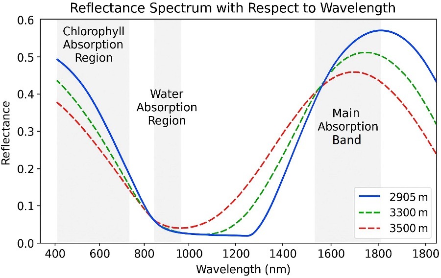
Рисунок 1. Спектры отражения листьев Quercus aquifolioides на разных высотах. Типичная кривая отражения растительности характеризуется сниженным отражением в видимом диапазоне вследствие поглощения пигментами, выраженным переходом «красного края» в ближнюю инфракрасную область с высоким NIR-отражением, обусловленным внутренней структурой листа, и отчётливой полосой поглощения воды около 1350–1450 нм. Затенённые области обозначают зоны поглощения хлорофиллом и водой, а основная полоса поглощения отражает структурные эффекты и влияние гидратации. Отражение около 1300 нм связано с динамикой оводнённости тканей листа (Zhu et al., 2020).
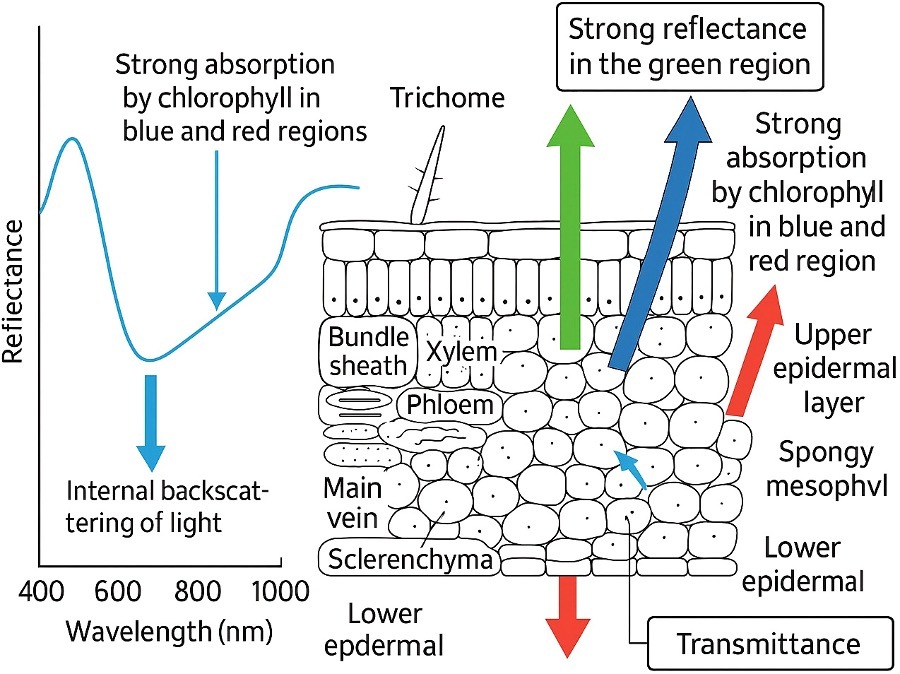
Рисунок 2. Поперечный срез типичного листа и соответствующий спектральный профиль отражения с обозначением типов и слоёв клеток, а также основных взаимодействий света при прохождении через эти структуры. Показаны сильное поглощение хлорофиллом в синей и красной областях спектра, сильное отражение в зелёной области, внутреннее обратное рассеяние в мезофилле и частичное пропускание через нижний эпидермис. Совокупность этих процессов объясняет характерный спектр отражения здоровой растительности (Liew et al., 2008).
1.1 Методология обзора
Для обеспечения полного и воспроизводимого охвата был применён структурированный рабочий процесс в стиле PRISMA, адаптированный к нарративному/обзорному исследованию оптического зондирования и машинного обучения для выявления стресса растений. По возможности сведения из отобранных исследований непосредственно включались в основной анализ и таблицы, чтобы визуальные элементы поддерживали изложение, а не прерывали его.
1.1.1 Базы данных и источники
Использовались Web of Science Core Collection, Scopus, PubMed, IEEE Xplore и Google Scholar (первые приблизительно 200 результатов по каждому запросу для ограничения информационного шума). Препринты, например arXiv, учитывались только при отсутствии рецензируемой версии.
1.1.2 Временной интервал и язык
Январь 2000 г. — апрель 2025 г.; только англоязычные публикации.
1.1.3 Критерии включения
Включались рецензируемые эмпирические исследования, в которых сообщалось: (i) об оптическом зондировании/визуализации растений от листа до полога и (ii) об алгоритмическом анализе — классическом или глубоком машинном обучении — либо о методических нововведениях, обеспечивающих выявление или количественную оценку стресса. Допускались исследования в теплицах и полевых условиях.
1.1.4 Критерии исключения
Исключались исследования нерастительных объектов или исключительно неоптических методов; неанглоязычные работы; редакционные статьи и комментарии; тезисы конференций без полного текста; диссертации; дубликаты; обзоры (их использовали только как фоновые источники); исследования без ключевых методических сведений, например без указания спектральных полос или схемы валидации.
1.1.5 Скрининг и отбор
После удаления дубликатов записи независимо проверяли два рецензента (MS, SUK) в два этапа: (1) название и аннотация; (2) полный текст. Разногласия разрешались при обсуждении с третьим автором (AQ). Поиск назад и вперёд по цитированию позволил выявить дополнительные подходящие работы. Причины исключения на этапе полного текста фиксировались, например недостаточно надёжная эталонная разметка или использование неоптических датчиков. Это обеспечило соответствие материалов, упомянутых на рисунках 1–15 и в таблицах 1–6, единым методическим требованиям. Применение визуализации на базе смартфона для оценки окраски листьев риса и выявления дефицита азота показано на рисунке 5 (Casari et al., 2019).
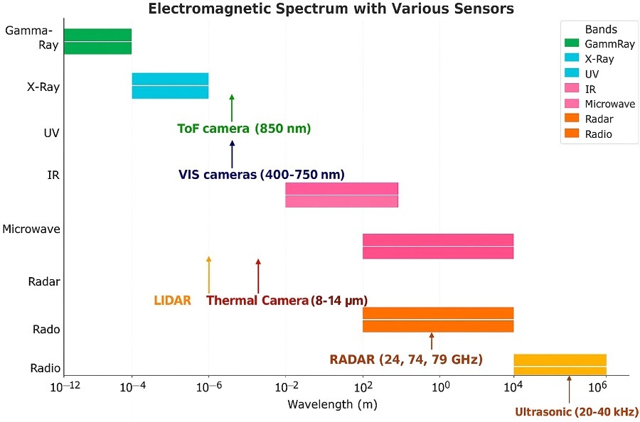
Рисунок 3. Участки электромагнитного спектра, используемые различными оптическими датчиками, включая ультрафиолетовый, видимый и ближний инфракрасный диапазоны, применяемые для выявления стресса растений. Адаптировано по Rosique et al. (2019).
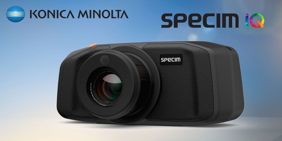
Рисунок 4. Компактная гиперспектральная камера Specim IQ. Адаптировано по Behmann et al. (2018).
1.1.6 Извлечение данных
Для каждого исследования регистрировали вид растения/культуру; тип стресса — биотический или абиотический; условия — теплица или поле; датчик/модальность и спектральные полосы; платформу — ручную, смартфонную или БПЛА; объём данных; эталонную разметку; предварительную обработку; метод обучения и протокол валидации; показатели качества; доступность кода и данных; основные ограничения. Эти сведения лежат в основе сравнительных материалов разделов 3–4, включая характеристики модальностей, индексы, компромиссы методов машинного обучения в таблице 4a и сводные результаты в таблице 6.
1.1.7 Оценка качества и соответствия назначению
Хотя метаанализ не проводился, была использована система оценки прозрачности и риска систематической ошибки по следующим направлениям: (i) выборка и повторности; (ii) строгость эталонных измерений; (iii) схема валидации — перекрёстная, на отложенной выборке либо на независимом участке/годе; (iv) работа с дисбалансом классов; (v) полнота отчётности — матрица ошибок и доверительные интервалы; (vi) воспроизводимость — доступность кода и данных. Оценки учитывались при определении веса результатов в повествовательном анализе. Они отражены при обсуждении ограничений и эталонных тестов в разделах 4.2.4 и 5.1.
1.1.8 Методические ограничения
Неоднородность конфигураций датчиков, условий и способов представления результатов ограничила возможность количественного синтеза. Хотя Google Scholar способен вносить информационный шум, использование нескольких баз данных и поиск по цепочкам цитирования снижали риск пропусков. Для связности изложения перекрёстные ссылки на рисунки и таблицы направляют читателя по ходу анализа; далее раздел 2 излагает спектральные основы.
1.1.9 Политика актуальности
Учитывая быстрый прогресс области, в качестве основных доказательств приоритет отдавался исследованиям 2020–2025 гг.; более ранние ссылки заменялись новыми, когда были доступны обновлённые работы, но основополагающие публикации сохранялись для исторического контекста. Этот подход отражён в разделах 3–4, например Patel et al. (2024a), Li et al. (2025a), Huang et al. (2023), Lei et al. (2024), Ammar et al. (2024), Kim et al. (2024), Zhang et al. (2023c), Liu et al. (2024b), Wang et al. (2025), Garcia et al. (2025a). Такая структура обеспечивает методическую согласованность рассмотренной литературы и повышает надёжность сравнительного анализа в разделах 3–5.
Таблица 1. Оптические методы выявления стресса растений
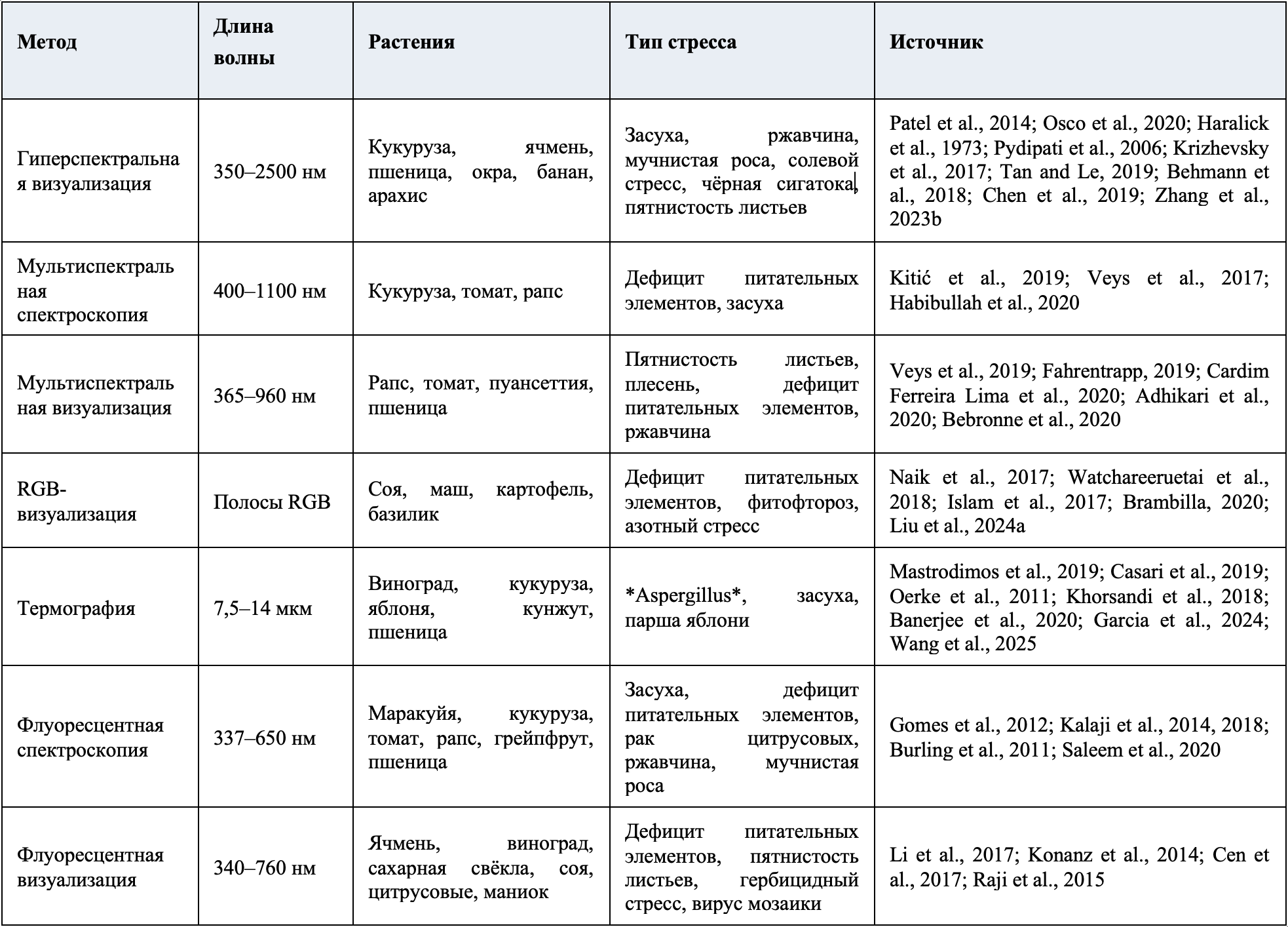
Таблица 2. Вегетационные и патологические индексы: уравнения и применение
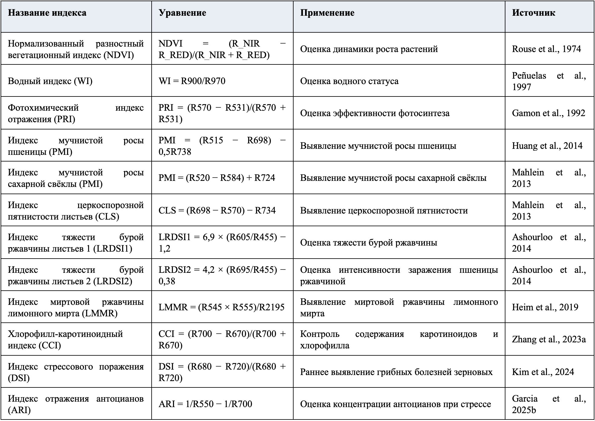
Таблица 3. Сравнительная оценка оптических подходов к мониторингу стресса растений
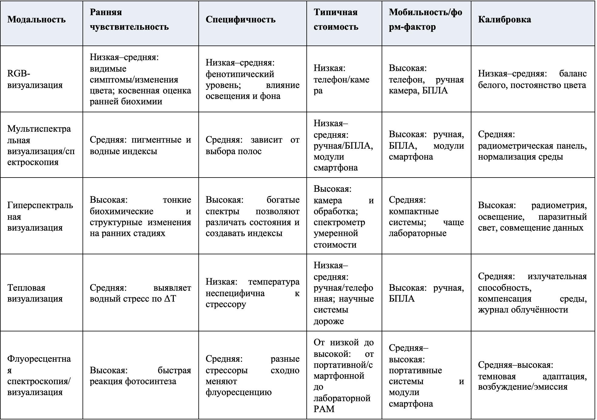
Примечание. Чувствительность отражает способность выявлять досимптомные или слабые сигналы; специфичность — способность различать стрессоры. Стоимость дана относительно разных модальностей и уровней оборудования. Пригодность предполагает соблюдение указанных правил калибровки.
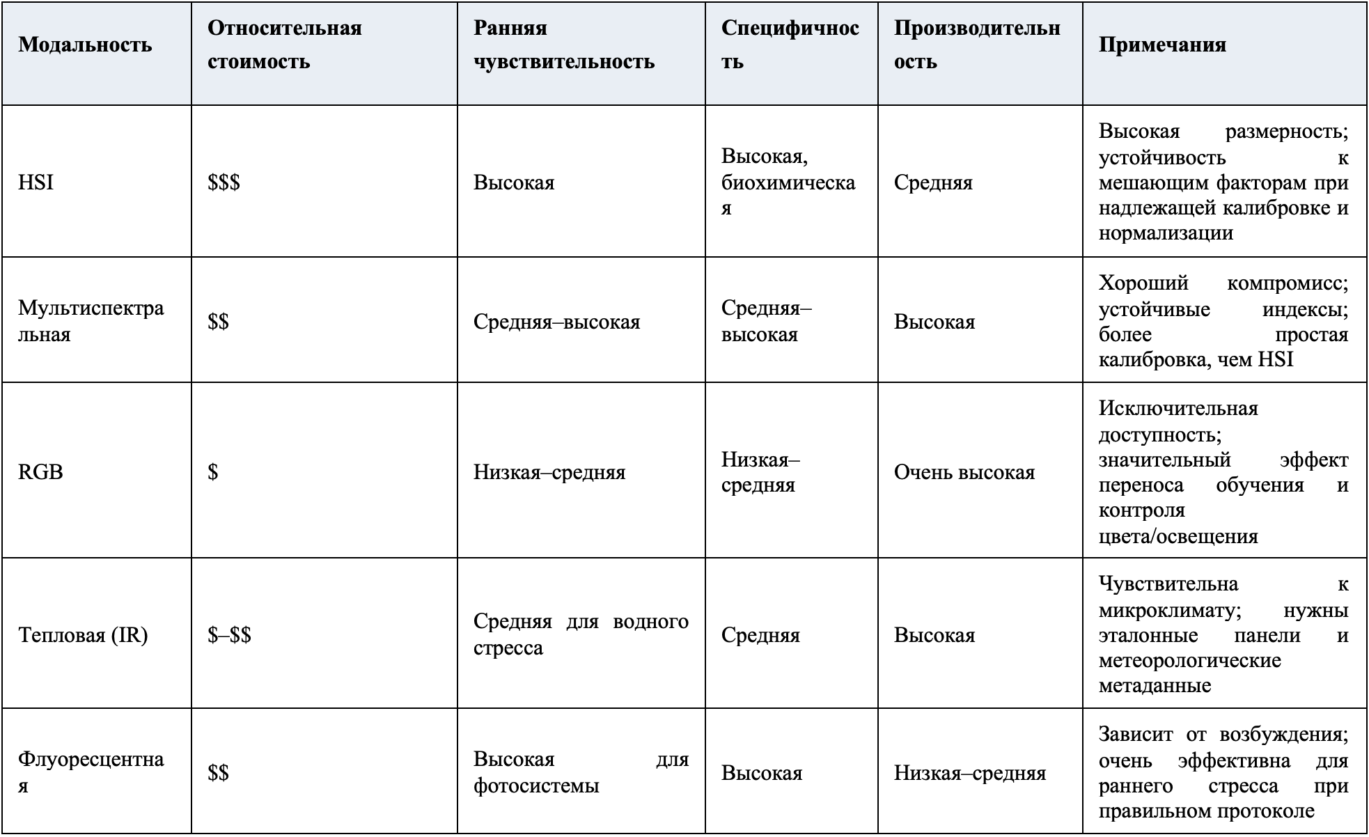
Таблица 4a. Сравнение модальностей визуализации для выявления стресса растений
Таблица 4a. Сравнение модальностей визуализации для выявления стресса растений
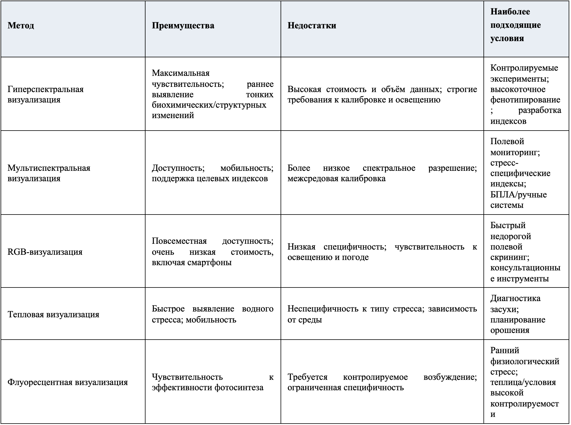
Таблица 4b. Сравнение подходов машинного обучения для выявления стресса растений
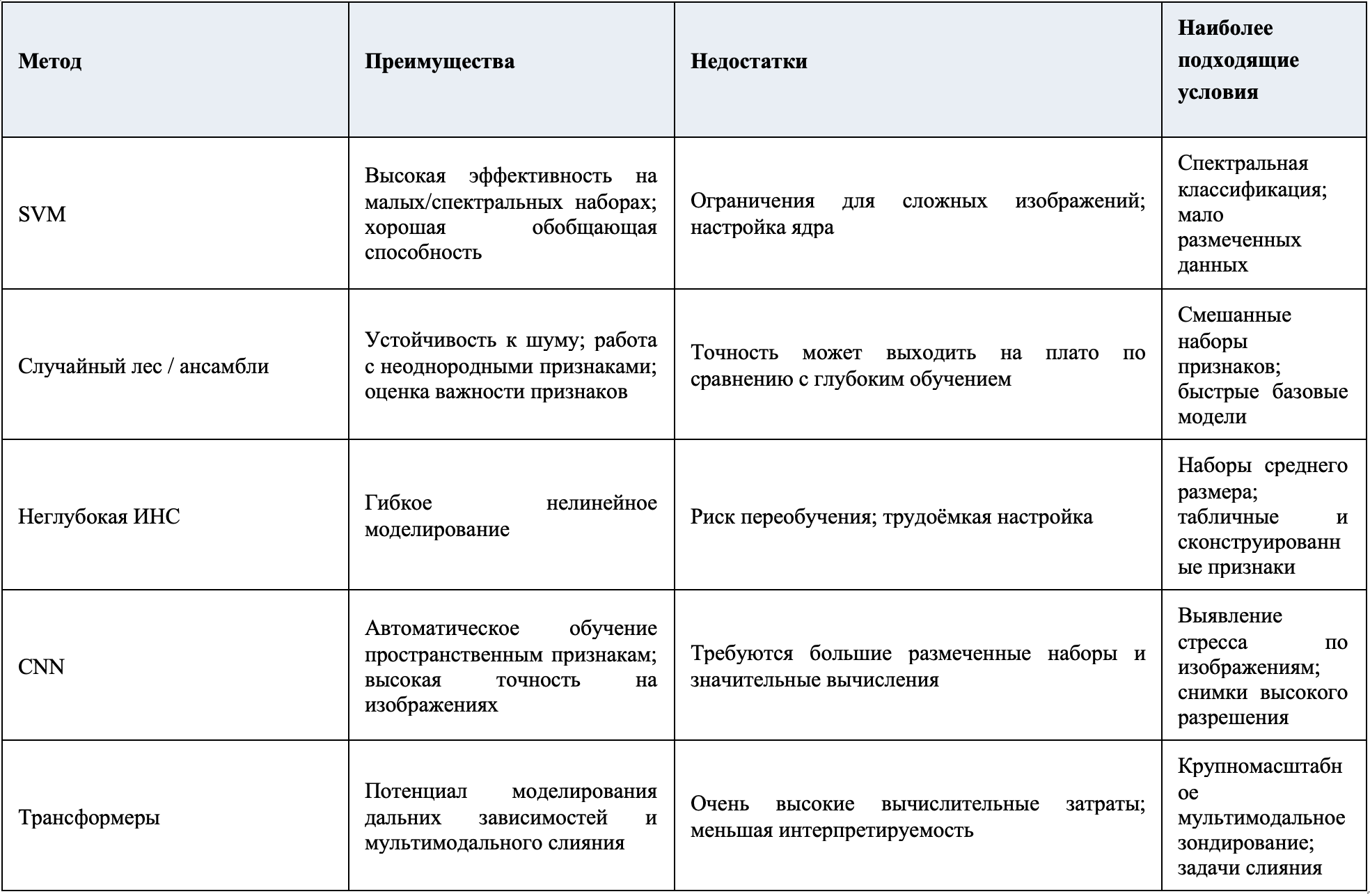
Таблица 5. Качественные компромиссы модальностей визуализации в полевых условиях
2 Спектральные свойства тканей растений
Спектральное отражение тканей растений определяется их физиологическими характеристиками и химическим составом, которые меняются под действием стресса (Singh et al., 2023). Такие изменения способны модифицировать профиль отражения листа, поэтому спектральный анализ полезен для диагностики (рисунок 1). Каноническая кривая растительности демонстрирует обусловленное пигментами поглощение в видимом диапазоне и выраженный «красный край» при переходе в NIR; при стрессе красный край обычно смещается, а поглощение в зелёной полосе уменьшается. Хлорофилл — ключевой пигмент фотосинтеза — особенно чувствителен к стрессу. Снижение его содержания обычно повышает отражение около 700 нм (Zhu et al., 2020) и одновременно уменьшает отражение в диапазоне 530–630 нм (Lichtenthaler et al., 1996; Gitelson etal., 2002). Эти изменения служат признаками нарушения эффективности фотосинтеза. Помимо хлорофилла, на отражение влияют другие пигменты (Vilfan et al., 2018; Li et al., 2022). Обусловленные пигментами сдвиги лежат в основе индексов PRI, CCI и ARI, сведённых в таблице 2 и далее используемых с конкретными датчиками в разделе 3.
Анатомические признаки листа (рисунок 2), включая форму клеток эпидермиса (Bone et al., 1985), шероховатость поверхности, толщину кутикулы (Grant et al., 1993) и плотность трихом (Ehleringer et al., 1976), также определяют спектральное поведение и нередко изменяются при стрессе. На рисунке 2 показаны эпидермис, палисадный и губчатый мезофилл; их толщина и архитектура внутренних воздушных пространств главным образом определяют поглощение видимого света и многократное рассеяние NIR, объясняя проявление структурных изменений в отражении. УФ-излучение, например, способно изменять содержание хлорофилла и толщину структур, влияя на флуоресцентное излучение (Bornmanand Vogelmann, 1991). Peñuelas et al. (1993) показали, что отражение в диапазоне 950–970 нм связано с эластичностью клеточной стенки, уменьшающейся при засухе. Устьица — микроскопические поры на поверхности листа — регулируют влажность и газообмен, но могут служить входными воротами для патогенов (Liew et al., 2008). Распознавая патоген-ассоциированные молекулярные паттерны, растение может закрывать устьица как защитную реакцию (Sawinski et al., 2013). Это ограничивает транспирацию и вызывает измеримое повышение температуры поверхности листа, регистрируемое инфракрасной визуализацией. Структурные и устьичные эффекты обосновывают применение теплового зондирования (раздел 3.4) и флуоресцентных методов (разделы 3.5–3.6), а таблица 2 содержит дополняющие индексы, например WI для водного статуса.
Разные стрессоры формируют характерные и часто диагностически значимые изменения профилей отражения. Засуха прежде всего ослабляет полосы поглощения воды около 1450 и 1950 нм вследствие обезвоживания, а также сдвигает красный край из-за усадки клеточных стенок и снижения рассеяния в мезофилле. Дефицит элементов питания, особенно азота и фосфора, уменьшает содержание хлорофилла и белка, снижая поглощение в видимой области и делая переход красного края более пологим. Солевой стресс нарушает целостность клеток мезофилла и изменяет углеводно-лигниновый состав, часто повышая отражение в SWIR-диапазоне 1300–2500 нм из-за ионного дисбаланса (de Lima et al., 2014). Патогенные инфекции косвенно изменяют отражение через перестройку поверхности листа, разрушение пигментов и локальный некроз, создавая неоднородные «пятна» отражения в видимой и NIR-областях (Mahlein et al., 2022). Такие мозаичные поражения ослабляют или смещают красный край и повышают локальную дисперсию в диапазоне 680–750 нм, формируя высокочастотную текстуру, которую можно использовать в признаках GLCM/энтропии или узкополосных индексах болезней PMI/CLS. Это повышает разделимость на гиперспектральных и мультиспектральных изображениях высокого разрешения. Эти стресс-специфические спектральные «отпечатки» непосредственно определяют выбор модальности в разделе 3 и диапазоны на рисунке 3.
Белки, целлюлоза, крахмал, лигнин, гемицеллюлоза и сахара также изменяются под воздействием среды и влияют на спектральный отклик (Fourty et al., 1996; Mahlein et al., 2022). Солевой стресс, например, способен нарушать целостность мезофилла и состав структурных углеводов — полисахаридов и лигнина (de Lima et al., 2014). Поскольку вода сильно поглощает свет за пределами 1300 нм в инфракрасном спектре, изменение её содержания — ещё одного критически важного маркера стресса — существенно влияет на спектральные характеристики (Allen and Richardson, 1968). Эти эффекты мотивируют применение датчиков 900–2500 нм (раздел 3.1) и ориентированных на воду индексов, таких как WI.
2.1 Перспективные направления исследований
Основная часть исследований сосредоточена на видимом и NIR-диапазонах, однако малоизученные области, включая SWIR1400–2500 нм и терагерцовый диапазон, перспективны для выявления биохимических маркеров — лигнина, вторичных метаболитов и стресс-индуцированных летучих соединений. Объединение спектральных измерений с омиксными данными, например метаболомикой и протеомикой, может выявить новые связи между биохимическими путями и спектральными признаками. Другое важное направление — универсальные спектральные биомаркеры, различающие перекрывающиеся реакции на стресс, что особенно важно в поле при одновременном действии нескольких стрессоров. Интеграция этих данных с моделями машинного обучения (раздел 4) может обеспечить более раннюю диагностику и ускорить перенос сенсорных технологий в точное земледелие. Механизмы, изложенные здесь, прямо определяют выбор датчиков и стратегию сбора данных в разделе 3, где спектральная чувствительность сопоставлена с практическими ручными, смартфонными и БПЛА-платформами, рабочими индексами (таблицы 1, 2) и пригодностью для поля (таблица 3; раздел 3.8), подготавливая анализ машинного обучения в разделе 4.
Описанные механизмы определяют, где в спектре находится диагностически полезный контраст и какие датчики наиболее информативны. Динамика хлорофилла и каротиноидов в видимой области и зоне красного края благоприятствует RGB- и VIS–NIR-мультиспектральным камерам и индексам PRI/CCI. Многократное рассеяние в мезофилле, повышающее NIR-отражение, обосновывает узкие полосы около красного края для структурно-чувствительных признаков. Содержание воды и эластичность клеточных стенок оставляют выраженные признаки после 1300 нм, что указывает на SWIR-гиперспектральные системы и водные индексы. Закрытие устьиц повышает температуру листа и обосновывает тепловую визуализацию. Изменения переноса энергии в фотосистемах регистрируются флуоресцентной спектроскопией и визуализацией. Раздел 3 переводит эти связи в практические платформы, полосы пропускания и полевые ограничения.
3 Датчики и сбор данных
На основе механизмов раздела 2 выбираются датчики, полосы пропускания которых пересекают диагностические признаки: видимые полосы и красный край — для пигментных и структурных сдвигов, SWIR — для воды и биохимического состава, тепловой IR — для реакций устьиц и энергетического баланса, а возбуждение и эмиссия флуоресценции — для кинетики фотосистем (таблица 1). Приборы регистрируют отражение, которое представляют изображениями или спектральными кривыми. Эффективность датчика определяется чувствительностью к областям профиля отражения, изменяемым биотическими и абиотическими факторами. Среди новых средств in situ — микроигольчатый пластырь для определения перекиси водорода в листьях, выявляющий ранние биохимические признаки до видимых симптомов (Dong et al., 2025). Видимый диапазон особенно информативен для оценки состояния растений (Carter, 1993), однако стресс проявляется и в других областях. pH-чувствительный датчик на основе хроматических ковалентных органических каркасов выявлял вызванные засухой изменения pH за 48 часов до визуальных симптомов (Strano et al., 2024). Рисунок 3 показывает положение модальностей в электромагнитном спектре, а таблица 1 — характерные длины волн, культуры и стрессоры; далее модальности связываются с индексами таблицы 2.
Платформа CropVoice использует генетически модифицированные растения, испускающие флуоресцентный сигнал при стрессе; его регистрируют датчики на дронах, спутниках или тракторах для мониторинга в реальном времени (Aronov et al., 2024). Такие биохимические репортёры дополняют признаки отражения и могут объединяться с оптическими измерениями для повышения специфичности раннего предупреждения.
3.1 Гиперспектральная визуализация
Гиперспектральная визуализация регистрирует тонкие пигментные, водные и структурные признаки во всём диапазоне VIS–NIR–SWIR, включая красный край и водные полосы выше 1300 нм, отмеченные в разделе 2. Она обеспечивает наиболее подробную биохимическую и структурную информацию для раннего выявления стресса, но имеет более высокую стоимость и сложность данных. Метод объединяет визуализацию и спектроскопию, создавая многомерные данные, где каждый пиксель подробно описан спектральными характеристиками на множестве отдельных длин волн (Pandey et al., 2017). Это позволяет точно различать пространственные области и тонкие спектральные различия, которые теряются при суммировании отражения по всему листу или изображению и могут смешивать здоровые и повреждённые ткани. Поэтому гиперспектральная визуализация стала мощным средством фенотипирования и скрининга стресса (Rumpf et al., 2010; Yanget al., 2019; Zovko et al., 2019). Она применялась для оценки реакции банана (Krizhevsky et al., 2017) и кукурузы (Patel et al., 2014) на засуху, жёлтой ржавчины пшеницы (Osco et al., 2020), стресса ячменя (Fourty et al., 1996), солевого стресса окры и мучнистой росы разных видов (Allen and Richardson, 1968).
Типичный диапазон гиперспектральных систем — 250–2500 нм, включая УФ и NIR; видимый и ближний инфракрасный диапазоны особенно важны для выявления стресса (Deng et al., 2009). Несмотря на большой объём работ по этим полосам, новые исследования осваивают другие участки спектра для получения новых индикаторов; например, недавно гиперспектральную визуализацию применили для оценки солевого стресса ячменя (Tan and Le, 2019).
Гиперспектральные данные часто используют через вегетационные индексы, рассчитываемые как отношения или разности отражения на определённых длинах волн (Saleem et al., 2019; Mohanty et al., 2016; Lu et al., 2017b). Наряду с ними создаются спектральные индексы болезней, ориентированные на конкретные патологии (Meng et al., 2020; таблица 2), например индексы мучнистой росы сахарной свёклы и пшеницы (Huang et al., 2014; Mahlein et al., 2013). Большой объём спектральной информации позволяет разрабатывать новые чувствительные к стрессу индексы для улучшения ранней диагностики (Peñuelas et al., 1997; Gamon et al., 1992).
Несмотря на устойчивость и информационную насыщенность, гиперспектральная визуализация традиционно ограничена стоимостью и размерами оборудования. Стандартные датчики громоздки и дороги, поэтому их трудно применять в реальном времени в поле. Технологическое развитие привело к портативным спектрорадиометрам и компактным камерам (рисунок 4); хотя их спектральный диапазон уже, они пригодны для целевой диагностики в реальном времени (Balasundramet al., 2020; Behmann et al., 2018). Спектрорадиометры не формируют гиперспектральные изображения, но успешно выявляли пятнистость листьев арахиса (Chen et al., 2019) и мучнистую росу ячменя (Behmann et al., 2018).
3.1.1 Ограничения и практические аспекты
При высокой диагностической способности метод имеет существенные ограничения. Системы часто стоят несколько тысяч долларов США, требуют тщательной радиометрической калибровки и контролируемого освещения, а большие массивы данных — сложных вычислительных конвейеров, что затрудняет полевую работу в реальном времени. Портативные спектрорадиометры и компактные камеры развиваются, однако более узкий спектральный охват ограничивает универсальность, а цена оборудования по-прежнему мешает широкому внедрению.
3.2 Мультиспектральная визуализация и спектроскопия
Мультиспектральные методы выбирают ключевые области спектра, прежде всего зелёную, красный край и полосы поглощения воды, переводя пигментные и структурные механизмы раздела 2 в практические каналы. Отбор нескольких диагностических полос вместо полного спектра снижает стоимость и упрощает развёртывание. В отличие от гиперспектральных методов, мультиспектральные подходы собирают данные лишь в нескольких диапазонах, а не в сотнях узких полос. Камеры и иные приборы формируют изображения отдельных областей — мультиспектральную визуализацию; мультиспектральная спектроскопия создаёт спектральные данные по заданным полосам.
Эти методы применялись для выявления пятнистости листьев рапса (Veys et al., 2019), серой гнили томата (Fahrentrapp, 2019), дефицита питания (Cardim Ferreira Lima et al., 2020), азотного стресса кукурузы (Kitić et al., 2019), засухи томата (Veys et al., 2017) и азотного стресса рапса (Habibullah et al., 2020). Они обычно дешевле гиперспектральных из-за широких полос, но дают меньше подробной информации о растении и среде. Высокая мобильность и универсальность делают их удобными для специализированных устройств: нужные полосы недорого получают с помощью полосовых фильтров и камер. Современные камеры смартфонов способны регистрировать NIR. Chung et al. (2018) применили съёмный высокочастотный фильтр 800 нм для одновременного получения красных и NIR-изображений. Смартфонные мультиспектральные устройства с глубоким обучением выявляли дефицит питательных элементов листовых овощей в реальном времени (Patel et al., 2024b), а дешёвые встроенные камеры подтверждены для диагностики засухи кукурузы (Li etal., 2025b). Новые интегрированные стеки с насадками и моделями на устройстве обеспечивают надёжную полевую диагностику питания и воды на малых участках (Patel et al., 2024b; Garcia et al., 2025a; Li et al., 2025b).
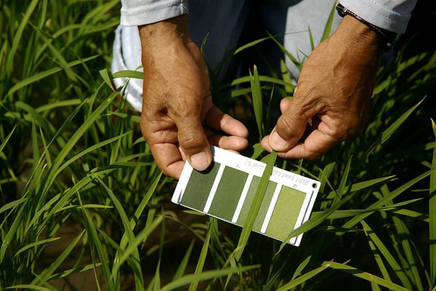
Рисунок 5. Применение смартфонной визуализации для оценки окраски листьев риса при выявлении дефицита азота. Показано полевое использование стандартной цветовой шкалы листьев для визуального сравнения степени зеленения, соответствующей азотному статусу. Повышение разрешения улучшает видимость градаций цвета и контекстных деталей (Casari et al., 2019).
3.2.1 Ограничения и практические аспекты
Мультиспектральные системы дешевле и мобильнее гиперспектральных, однако более низкое разрешение может скрывать тонкие физиологические сигналы. Межсредовая калибровка нетривиальна; качество зависит от фильтров и типа сенсора. Смартфонные устройства перспективны как недорогой вариант, но различия камер и прошивок усложняют стандартизацию. Предварительная обработка и индивидуальная калибровка устройств необходимы для устойчивых показателей, приведённых в таблице 6.
3.3 RGB-визуализация
RGB-визуализация полностью работает в видимом спектре и чувствительна к обусловленным пигментами вариациям отражения. Она позволяет создавать цветовые показатели, аналогичные PRI и CCI, которые косвенно характеризуют динамику хлорофилла и каротиноидов. Не регистрируя NIR и SWIR, RGB-сенсоры всё же обеспечивают дешёвый доступ к связанным со стрессом изменениям окраски. Видимый свет регистрируется в синей (400–499 нм), зелёной (500–549 нм) и красной (550–750 нм) полосах с максимумами отклика около 475, 520 и 650 нм соответственно (Deng et al., 2009). Поэтому RGB можно считать подмножеством мультиспектральной визуализации. Обычно применяются цифровые камеры и смартфоны, тогда как полноценная мультиспектральная съёмка требует специализированных приборов (Watchareeruetai etal., 2018).
Ключевые преимущества RGB — низкая стоимость и компактность сенсора. Поскольку смартфоны уже содержат RGB-камеры, они являются практичной и доступной платформой. Метод выявлял железодефицитный хлороз сои (Naik etal., 2017), недостаток элементов питания у маша (Xie et al., 2016), ранний и поздний фитофтороз картофеля и грибные болезни пшеницы (Islam et al., 2017). Повсеместность смартфонов и встроенные вычисления позволяют быстро анализировать данные без глубокой подготовки, расширяя доступ к мониторингу и полевой оценке в реальном времени (Taoet al., 2020; Naik et al., 2017; Chen et al., 2024; Li et al., 2025a). Пример для листьев риса показан на рисунке 5. RGB не чувствителен к SWIR, но косвенно отражает пигментную динамику индексов CCI/ARI; сочетание с обучаемыми признаками часто восстанавливает значительную долю диагностического сигнала при минимальной цене оборудования.
Качество RGB зависит от яркости, внешней среды, времени суток и спектрального отклика камеры (Mattupalli et al., 2018), особенно от сезонных и погодных изменений солнечного света. Для повышения точности используют постоянство цвета и нормализацию освещения с серой картой/балансом белого, удаление теней и сегментацию листа, затем перенос обучения и аугментацию. Индивидуальная калибровка устройств и простая адаптация домена между участками и камерами повышают устойчивость и поддерживают быстрый вывод на устройстве (Naik et al., 2017).
3.3.1 Ограничения и практические аспекты
Доступность RGB сочетается с высокой чувствительностью к освещённости, углу солнца, погоде и отклику сенсора, ухудшающим воспроизводимость. Различия между смартфонами и отдельными камерами дополнительно снижают надёжность. Метод не обладает прямой чувствительностью ко многим биохимическим маркерам и ограничен по специфичности. Наиболее эффективны развитая обработка/машинное обучение и сочетание с тепловыми, флуоресцентными или узкополосными данными. Таблица 4b рекомендует RF/SVM для малых RGB-наборов и CNN/трансформеры при большом объёме разметки.
3.4 Тепловая визуализация, или термография
Значимость тепловой визуализации непосредственно следует из механизмов устьичной регуляции и транспирации. При стрессе устьица закрываются, транспирация уменьшается, а поверхность листа нагревается. Тепловой IR регистрирует эту разность температур как прямой физиологический показатель водного стресса и энергетического баланса. В отличие от других методов термография измеряет испускаемое, а не отражённое излучение (Mastrodimos et al., 2019). Тепловые камеры представляют температуру ложными цветами, где каждому пикселю соответствует измеренное значение.
Изменение температуры листьев позволяет рано выявлять стресс. При дефиците воды закрытие устьиц уменьшает транспирацию и повышает температуру (Jones, 1999). Новые датчики и их интеграция с ИИ повысили чувствительность и качество диагностики (Garcia et al., 2024; Wang et al., 2025). Метод применяли для выявления Aspergillus carbonarius на винограде (Oerke et al., 2011), парши яблони (Casari et al., 2019), засухи кукурузы и кунжута (Jones, 1999; Khorsandi et al., 2018). Рисунок 6 показывает разделение водных режимов кукурузы по тепловому контрасту. Этот контраст прямо отражает закрытие устьиц, однако неспецифичен; для установления причины обычно необходимо слияние со спектральными/флуоресцентными данными и обучаемыми правилами.
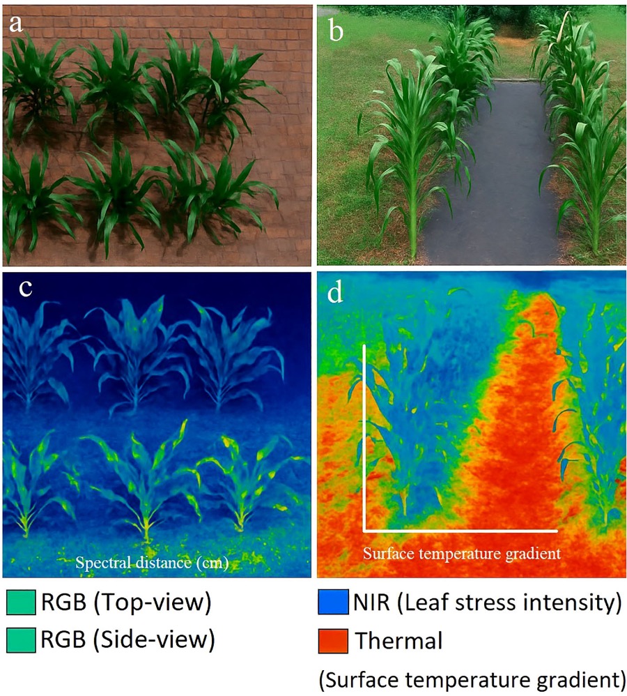
Рисунок 6. Мультимодальная визуализация кукурузы при засухе и достаточном поливе. (a) RGB-вид сверху и (b) RGB-вид сбоку показывают морфологические различия. (c) NIR-флуоресцентное изображение выявляет различия интенсивности стресса листьев; (d) тепловое изображение показывает градиенты температуры поверхности полога (°C), где более тёплые цвета соответствуют более сильному стрессу. На панелях приведены масштабные линейки и калиброванные легенды. Верхний ряд — достаточный полив; нижний — засуха. Перепечатано с разрешения Casari et al. (2019).
Термография проста и быстра. Портативные камеры, включая насадки для смартфонов, сделали её доступнее (Petrie et al., 2019). Однако результат сильно зависит от среды, что ограничивает надёжность в поле. Кроме того, метод не различает типы стресса, поэтому наиболее эффективен в сочетании с дополнительной диагностикой (Oerke et al., 2011; Khorsandi et al., 2018).
3.4.1 Ограничения и практические аспекты
Температура листьев сильно искажается ветром, влажностью, облучённостью и структурой полога. Хотя портативные камеры всё доступнее, приборы высокого разрешения и чувствительности остаются умеренно дорогими. Тепловой контраст неспецифичен: одной термографии недостаточно для надёжного определения стрессора, поэтому требуются мультимодальное слияние и контекстные данные.
3.5 Флуоресцентная спектроскопия
Флуоресцентная спектроскопия оценивает активность фотосистем по кинетике возбуждения и эмиссии хлорофилла и связанных пигментов. Стресс изменяет интенсивность и отношения флуоресценции, формируя ранние биохимические показатели эффективности фотосинтеза и дополняя измерение отражения. Спектрофотометрическая визуализация регистрирует ослабление падающего света при прохождении через листья, но изображения сложны из-за перекрывающихся спектров многочисленных пигментов и соединений (Lang et al., 1992). Флуоресцентные методы более избирательны, поскольку флуоресцирует ограниченное число компонентов: они поглощают коротковолновый и испускают более длинноволновый свет, что разделяет возбуждение и эмиссию (Lang et al., 1992; Krause and Weis, 1984). Метод выявляет снижение фотосинтетической активности при патогенном стрессе (Swarbrick et al., 2006).
Флуоресценция с импульсно-амплитудной модуляцией (PAM) широко применяется для оценки эффективности фотосинтеза. Используются импульсные и насыщающие источники вместе с непрерывным актиничным светом (Lawson and Vialet-Chabrand, 2018). Кинетика хлорофилльной флуоресценции во времени показывает влияние среды на фотосинтез (Brooks andNiyogi, 2011). При стрессе фотосинтетическая эффективность падает, а флуоресценция усиливается вследствие рассеяния энергии. Для точной кинетики обычно требуется 30-минутная темновая адаптация, позволяющая установить минимальную базовую флуоресценцию перед возбуждением (Lei et al., 2016; Lichtenthaler and Rinderle, 1988; Murchie and Lawson, 2013); необходима и адаптация к свету (Gomes et al., 2012; Kalaji et al., 2014, 2018).
Для оценки стресса используют отношения F440/F690, F440/F740 и F690/F740, особенно при УФ-возбуждении 320–400 нм (Buschmann and Lichtenthaler, 1998). Burling et al. (2011) показали, что отношения амплитуд красной/дальней красной и синей/зелёной флуоресценции различают дефицит азота и грибную инфекцию. Эти отношения можно дополнительно оптимизировать; воспроизводимость требует стандартизации темновой адаптации и возбуждения. Метод позволяет локализовать и количественно оценивать отдельные компоненты листа. Спектры табака при возбуждении 488 нм показаны на рисунке 7 (Lei et al., 2016).
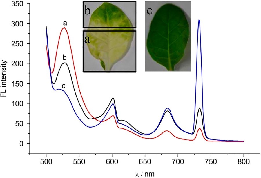
Рисунок 7. Спектры флуоресцентной эмиссии листьев табака при возбуждении 488 нм: (a) хлороз при заражении вирусом мозаики огурца (CMV); (b) заражённый CMV лист нормальной зелёной окраски; (c) здоровый контроль (Lei et al., 2016).
Флуоресцентная спектроскопия обеспечивает недорогую ручную диагностику засухи кукурузы и маракуйи (Gomes et al., 2012), дефицита питания кукурузы и томата (Kalaji et al., 2014) и рака цитрусовых у грейпфрута (Murchie and Lawson, 2013). Лазерное возбуждение повышает чувствительность, но изменения флуоресценции могут иметь разные причины, поэтому для специфичной диагностики метод следует дополнять другими средствами (Saleem et al., 2020). При кинетических исследованиях интенсивность со временем снижается; Saleem et al. (2020) уменьшили влияние этого эффекта, анализируя спектр в течение 15 секунд после возбуждения.
3.5.1 Ограничения и практические аспекты
Высокая чувствительность к нарушению фотосинтеза сочетается с потребностью в темновой адаптации или контролируемом освещении и временным затуханием сигнала. Специфичность ограничена, поскольку разные стрессоры дают сходные изменения. Оборудование варьирует от дешёвого портативного до лабораторного, что создаёт проблемы стоимости и масштабирования. Быстрая регистрация частично помогает, но массовое полевое применение требует стандартизации. Сочетание с RGB, тепловыми или узкополосными данными и машинным обучением повышает качество ранней диагностики и установления причины.
3.6 Флуоресцентная визуализация
Флуоресцентная визуализация отображает пространственно распределённую эмиссию и неоднородность листьев/пологов. Карты красной и дальней красной флуоресценции локализуют реакции, связанные с нарушением фотосинтеза, и дополняют RGB и тепловую визуализацию. Камеры регистрируют пространственную флуоресценцию (рисунок 8), поэтому метод часто превосходит спектроскопию по размерности данных (Su et al., 2019). Если спектроскопия собирает единственную спектральную сигнатуру выбранной области, визуализация разделяет интересующие участки и фон по пространственной интенсивности (Konanz et al., 2014). Многоцветная визуализация с УФ-возбуждением регистрирует синюю F440, зелёную F520, красную F680 и дальнюю красную F740 полосы и объединяет их в составные изображения.
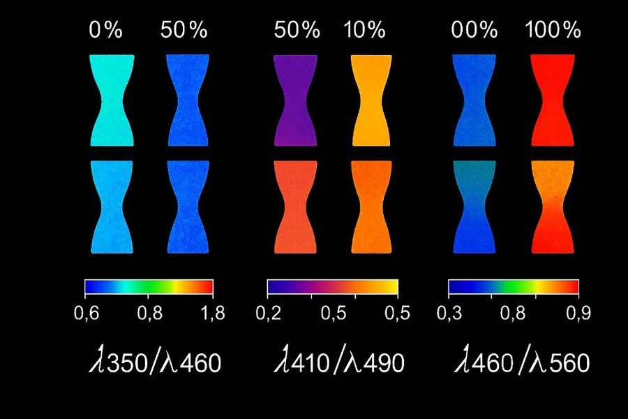
Рисунок 8. Отношения флуоресценции листьев ячменя при возрастающей степени дефицита азота. Перепечатано по Konanzet al. (2014).
Подобно мультиспектральной съёмке, метод выявляет биотические и абиотические стрессы: гербицидный стресс сои (Li etal., 2017), холодовой стресс рассады томата (Dong et al., 2019), стресс винограда, ячменя и сахарной свёклы (Su et al., 2019). Портативные системы, включая смартфоны с полосовыми фильтрами, потенциально пригодны для ранней диагностики, хотя исследованы недостаточно (Chung et al., 2019). Новые модули со смартфонным управлением, фильтрами и контролируемым УФ-возбуждением стали полевыми устройствами раннего скрининга и картирования листьев (Garcia et al., 2025b). При сравнительно низкой стоимости метод иногда не различает здоровую и больную ткань в начале инфекции, поэтому нуждается в дополнительной диагностике (Pérez-Bueno et al., 2019). Его важное преимущество — высокая чувствительность и возможность различать некоторые стрессоры (Takayama and Nishina, 2009). Пространственный контекст помогает машинному обучению отделять поражения от фона и нередко улучшает раннее выявление без существенного удорожания.
3.6.1 Ограничения и практические аспекты
Даже при невысокой стоимости оборудования метод может быть недостаточно эффективен в самом начале инфекции и чувствителен к схеме возбуждения/эмиссии и фоновой флуоресценции. Смартфонные реализации развиваются, но их устойчивость в разнообразных полевых условиях ещё предстоит подтвердить. Объединение с другими потоками данных повышает специфичность.
3.7 Сочетание датчиков
Для полной и надёжной оценки состояния растений рекомендуется объединять несколько методов. Слияние данных разных датчиков повышает точность и устойчивость, снижая влияние изменений среды (Moshou et al., 2011), однако согласование и интерпретация неоднородных потоков сложны. Berdugo et al. (2014) объединили тепловую, гиперспектральную и хлорофилл-флуоресцентную информацию дискриминантным анализом. Это позволило точно диагностировать мучнистую росу и различить огурцы, заражённые CMV и вирусом зелёной крапчатой мозаики. Дополняющие датчики повышают специфичность, но крупномасштабные данные разных структур требуют мощных аналитических и вычислительных методов.
Машинное обучение эффективно интерпретирует высокоразмерные данные мультимодального слияния и моделирует сложные нелинейные связи. Разные оптические датчики уже используются для оценки стресса культур (Adhikari et al., 2020; Bebronne et al., 2020; Brambilla, 2020; Banerjee et al., 2020; Cen et al., 2017; Raji et al., 2015), а развитые алгоритмы способны значительно усилить их эффективность. В обзоре индексы таблицы 2 сопоставлены с модальностями, показывая взаимодополняющую чувствительность, например SWIR-полосы воды + тепловая температура + флуоресцентный PRI. Эти сочетания определяют матрицу решений и выбор классификатора в разделе 4.
3.8 Сравнительная оценка подходов мониторинга
Таблица 3 сравнивает основные модальности по практическим критериям полевого внедрения: чувствительности и специфичности, стоимости, мобильности, калибровке и пригодности. Единственного «лучшего» метода нет: выбор определяется балансом раннего выявления, специфичности и цены. Гиперспектральные и флуоресцентные системы лучше выявляют ранние физиологические изменения; RGB и мультиспектральные методы предпочтительны для масштабируемого охвата; тепловая визуализация быстро сигнализирует о водном стрессе. Переносимость результатов определяется радиометрической калибровкой, журналами освещения, коррекцией излучательной способности и среды для тепловых датчиков, темновой адаптацией и контролем возбуждения для флуоресценции. В недорогих системах обычно применяют RGB/мультиспектральные данные и индексы с RF или SVM; в высокоточном фенотипировании — HSI с CNN или трансформерами. Тепловая съёмка вместе с RGB/мультиспектральной поддерживает управление орошением и установление причины стресса.
3.9 Сравнительная матрица выбора датчиков и алгоритмов
Таблицы 4a и 4b сопоставляют модальности и методы обучения по эффективности, цене/вычислениям, мобильности, масштабируемости и оптимальным сценариям. Панель (a) связывает физику модальности с ограничениями внедрения; панель (b) — свойства данных с семействами алгоритмов.
Гиперспектральные системы наиболее чувствительны к тонким биохимическим и структурным изменениям, но дороги и вычислительно интенсивны. Мультиспектральные методы балансируют информативность, доступность и мобильность и подходят для целевых индексов. RGB повсеместна и дешева, но ограничена по специфичности и зависит от среды. Тепловая визуализация быстро выявляет температурные изменения при водном стрессе, однако не различает стрессоры. Флуоресценция хорошо оценивает эффективность фотосинтеза, но часто требует контролируемых условий. Поэтому датчик следует выбирать по задаче, ресурсам и требуемой точности; мультимодальное слияние уменьшает ограничения отдельных методов.
Аналогичные компромиссы существуют между алгоритмами. SVM и RF эффективны для малых спектральных наборов, требуют умеренных вычислений, а RF оценивает важность признаков, однако эти методы хуже работают со сложной структурой изображений. ИНС моделируют нелинейности, но переобучаются без регуляризации. CNN и трансформеры достигают передовых результатов и моделируют пространственные/дальние зависимости, однако требуют больших размеченных наборов, высоких вычислений и менее интерпретируемы. Ансамбли и гибриды предлагают компромисс между пиковой точностью, масштабируемостью и интерпретируемостью. Решение зависит от объёма и типа данных, вычислительного бюджета и цели — скрининга или поддержки решений.
3.10 Синтез проектных шаблонов
Баланс раннего выявления и специфичности центральный для выбора сенсора. Флуоресценция и HSI обнаруживают ранние физиологические сдвиги; тепловая съёмка быстро выявляет водный стресс, но неспецифична; RGB и мультиспектральная съёмка обеспечивают широкий пространственный охват. Выбор определяется положением задачи в треугольнике «ранность–специфичность–стоимость».
Лестница слияния начинается с позднего слияния решений, наиболее устойчивого к отсутствующим данным, и переходит к слиянию признаков при надёжном совмещении и контроле освещения. Слияние на уровне исходных данных следует применять только при строгой стандартизации выравнивания и регистрации. Рекомендуется сообщать результаты абляционных исследований и устойчивость к пропуску модальности.
Сопоставимую полезность можно получить разными путями: объединить RGB/мультиспектральные данные с индексами и RF/SVM в недорогой маловычислительной системе либо использовать HSI с CNN/трансформерами для большей чувствительности ценой вычислений. Усиление метода оправдано только тогда, когда дополнительная чувствительность имеет операционное значение. Классические SVM и RF сильны на малых тщательно подготовленных спектральных наборах; глубокое обучение выгоднее при большом числе меток или мультимодальных входах. Для переносимости важнее межучастковая и многолетняя валидация, нормализация освещения и журналы калибровки, чем архитектура сама по себе.
Для малых хозяйств подходят RGB/мультиспектральные датчики с RF/SVM и индексами, возможно с тепловым каналом. Крупные предприятия используют БПЛА с мультиспектральными/тепловыми камерами и слияние признаков в системах поддержки решений. В исследовательском фенотипировании предпочтительны HSI/флуоресценция с CNN/трансформерами. Для воспроизводимости следует сообщать метаданные съёмки, калибровку, схему разделения без утечки, метрики по классам и калибровку вероятностей, абляции и слияние, стоимость на гектар и время до предупреждения.
У каждой модальности свой профиль стоимость–чувствительность–специфичность. Смартфоны добавляют вариабельность освещения, обработку прошивки и неоднородность устройств — автоматический баланс белого, резкость и тональное отображение. Для сопоставимости следует: (i) по возможности снимать RAW; (ii) фиксировать экспозицию, ISO и баланс белого; (iii) помещать в кадр серую/отражательную карту или Spectralon; (iv) стандартизировать расстояние и угол простыми направляющими; (v) регистрировать облучённость, ветер и температуру; (vi) применять цветовые профили конкретного устройства. Эти меры уменьшают дрейф и повышают переносимость вместе с переносом обучения и адаптацией домена (Patel et al., 2024a; Li et al., 2025a; Li et al., 2023).
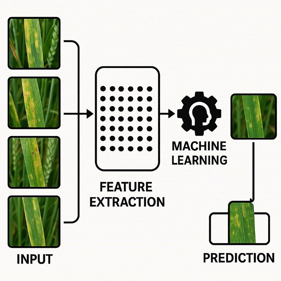
Рисунок 9. Базовый конвейер анализа данных машинного обучения: сбор данных, выделение признаков и классификация (Liakos et al., 2018).
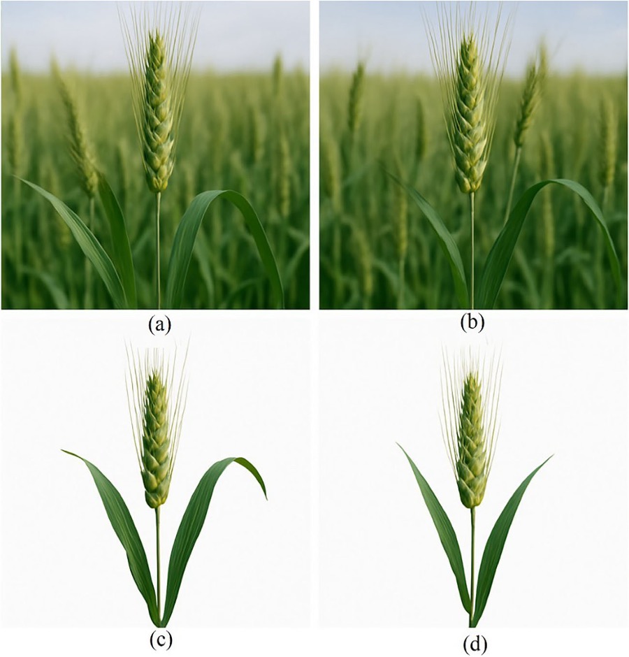
Рисунок 10. Визуализация сегментации. Исходные изображения хорошо орошаемой и испытывающей засуху пшеницы. Сначала значения RGB и линейная SVM использованы для первичной сегментации (a, b), затем математическая морфология — для удаления шума (c, d) (Zhuang et al., 2017).
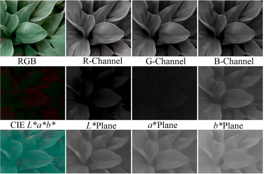
Рисунок 11. Другие цветовые пространства, полученные из RGB-изображения (цветового спектра) (Kahu et al., 2018).
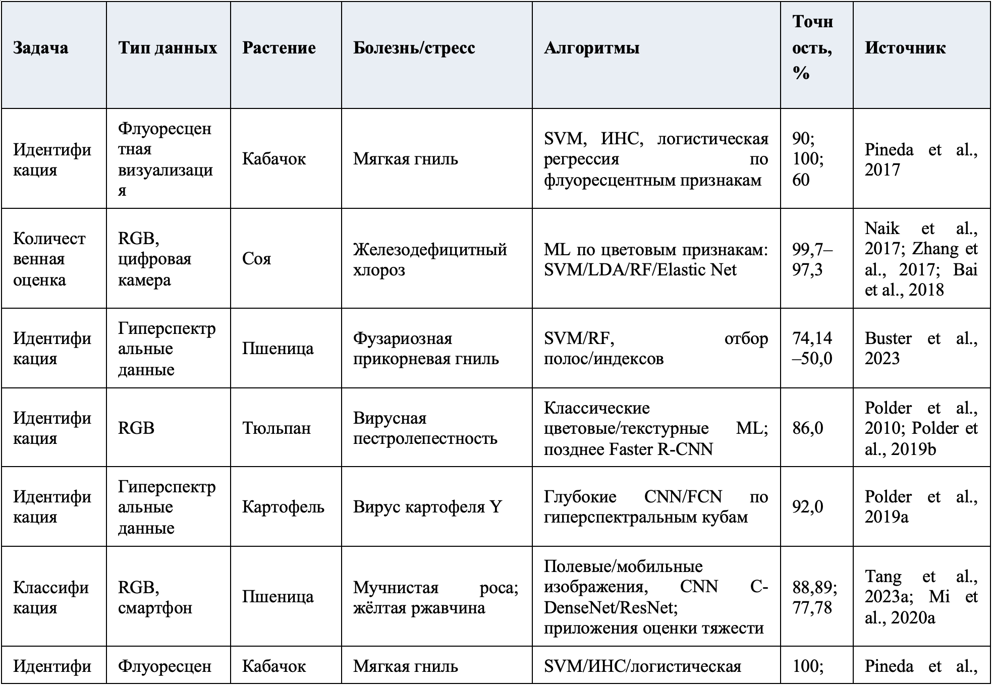
Таблица 6. Подходы ML/DL к выявлению и количественной оценке болезней/стрессов растений по различным типам изображений
4 Машинное обучение для анализа данных мультимодальных датчиков
Машинное обучение открыло новые возможности обработки данных в медицине, экологии, экономике и других областях. В простейшем виде алгоритмы обучаются на существующих данных без явного программирования каждого правила (Singh etal., 2016) и обнаруживают закономерности, пропускаемые традиционным анализом. Типичный процесс включает сбор и хранение данных, предварительную обработку, выделение признаков и классификацию (Ramos-Giraldo et al., 2020; рисунок 9). Раздел связывает типы данных и ограничения внедрения с семействами алгоритмов (таблица 4b), а результаты — с таблицей 6, учитывая индексы таблицы 2 и шаблоны раздела 3.10.
ML всё чаще используется для мультимодальных данных стресса растений, объединяя спектральные и тепловые признаки разных культур и условий. Ансамблевые и ядерные методы хорошо моделируют нелинейные связи между спектральными признаками и факторами среды и применяются для прогнозирования засухи, оптимизации питания и адаптивного выбора культур в вододефицитных районах (Farooqui and Ritika, 2019; Adhikari et al., 2020; Banerjee et al., 2020). SVM и случайный лес широко используются для спектральной классификации благодаря хорошему обобщению при ограниченной разметке.
В сельском хозяйстве ценность ML связана со способностью одновременно анализировать множество взаимодействующих переменных (Singh et al., 2016). Алгоритмы управляют сложностью через классификацию, снижение размерности и выделение признаков. Оценка состояния растений включает идентификацию, количественную оценку и различение. Идентификация распознаёт стрессор и отделяет его симптомы от других; количественная оценка измеряет интенсивность. Результаты приведены в таблице 6. Быстрая схема выбора: спектральные векторы с малым числом меток — SVM/RF; изображения высокого разрешения или мультимодальное слияние — CNN/трансформеры. Универсального метода нет; далее рассмотрены основные подходы.
4.1 Предварительная обработка
Подготовка данных необходима для точности и повторяемости классификации (Tsaftaris et al., 2016). Она стандартизирует вход и делает его доступнее алгоритму. Для изображений применяют кадрирование, удаление фона, усиление контраста, пороговую обработку, фильтрацию и кластеризацию (Singh et al., 2016). Сегментация хорошо орошаемой и испытывающей засуху пшеницы показана на рисунке 10. PCA также применяется к спектрам для снижения размерности и фильтрации шума.
Современные конвейеры объединяют нормализацию спектров — тёмную/белую коррекцию, сглаживание Савицкого–Голея, производные, SNV/MSC — и отбор полос с глубокими экстракторами, например EfficientNet или лёгкими автоэнкодерами (Patel et al., 2024a). При малом числе меток помогают самоконтролируемое предобучение и целевая аугментация. Обработка должна учитывать свойства сенсора: контроль возбуждения для флуоресценции; радиометрическую нормализацию HSI; постоянство цвета RGB/мультиспектральных данных; тёмную/белую коррекцию, виньетирование и паразитный свет; излучательную способность и средовую компенсацию теплового канала; спектральную «улыбку» и выравнивание полос HSI; цветовую карту/ICC-профиль и линеаризацию гаммы RGB; точное геометрическое совмещение мультимодальных данных и журнал метаданных съёмки.
4.1.1 Преобразование цветового пространства
Преобразование цветового пространства переводит RGB в другие представления для улучшения анализа и извлекает дополнительные цветовые атрибуты. Ручные дескрипторы обычно сочетаются с SVM/RF на малых наборах, тогда как глубокие модели способны сами обучаться инвариантности цвета. Пространство Lab* (L — светлота, a — ось зелёный–красный, b — ось синий–жёлтый) использовалось для выявления гнили плодов, бактериального ожога, пятнистости листьев и плодов граната (Dhakate and Ingole, 2015). HSI — тон, насыщенность, интенсивность — применялось для позднего и раннего ожога, мелкой белой пятнистости, хлопчатой и пепельной плесени (Al Bashish et al., 2010). YCbCr, где Y — яркость, Cb/Cr — сине- и красноразностные компоненты, использовали для болезней сои (Shrivastava et al., 2015). Новые преобразования повышают точность диагностики разных культур (Wei et al., 2024); пример показан на рисунке 11 (Kahu etal., 2018).
4.1.2 Снижение размерности
Снижение размерности упрощает данные при сохранении большей части информации. Анализ главных компонент (PCA) проецирует набор в пространство меньшей размерности (Lever et al., 2017), объединяя коррелированные переменные с минимальной потерей. PC1 объясняет наибольшую дисперсию, затем ортогональные PC2, PC3 и т. д. с убывающей долей. Компоненты визуализируют на двумерных/трёхмерных графиках и подают в ML-модели. PCA широко применяется к спектрам и изображениям; Lu et al. (2017a, 2017b) использовали его для вычисления карт признаков. Метод остаётся непредвзятым и распространённым, но чувствителен к выбросам (Wold et al., 1987). Сочетание PCA с другими методами улучшает выделение признаков в сложных наборах (Lei et al., 2024). Для малых спектральных данных типична лёгкая схема PCA + SVM/RF.
4.1.3 Сегментация
Сегментация делит изображение на значимые части — объект интереса и фон — и уменьшает ошибки из-за фонового шума. Кластеризация k-средних успешно применялась для распознавания стресса (Al Bashish et al., 2010); используются также удаление пикселей и маскирование (Singh et al., 2016; Ma et al., 2018). Новые U-Net и YOLOv8-Seg точнее очерчивают поражённые области (Ammar et al., 2024). Сегментация улучшает измерение поражений и уменьшает ложные корреляции, что отражается в более высоких precision/recall в таблице 6.
4.1.4 Выделение признаков
Выделение признаков преобразует сырой материал в удобное представление для алгоритмов, удаляя избыточность и сохраняя значимые свойства (Tsaftaris et al., 2016). Применяются локальные бинарные шаблоны LBP (Yue et al., 2011), вектор согласованности цвета CCV (Ojala et al., 2002), глобальная цветовая гистограмма GCH (Vatamanu et al., 2013), дисперсия каналов, однородность и контраст текстуры (Ma et al., 2018). Затем классификатор принимает решение по изученным закономерностям. Глубокие модели повысили точность диагностики (Patel et al., 2024a), однако вручную созданные признаки можно точнее адаптировать к модальностям таблицы 1.
4.2 Подходы машинного обучения к классификации
После подготовки набор передаётся алгоритму, который обнаруживает закономерности и классифицирует ранее неразмеченные объекты, например стрессовые и здоровые растения (Rumpf et al., 2010). Различают обучение с учителем, без учителя и со слабым учителем (Wang et al., 2017; Zhou, 2018). Первое требует размеченных данных; слабый контроль использует ограниченные, шумные или грубые метки; обучение без учителя работает без меток. Пример последнего — кластеризация сходных образцов (Rodriguez et al., 2019). Самоконтролируемые и гибридные методы помогают обрабатывать неструктурированные аграрные данные (Huang et al., 2023). Линейные и нелинейные границы и семейства моделей показаны на рисунках 12, 13; внимание и атрибуции — на рисунках 14, 15.
В сельском хозяйстве широко применяются искусственные нейронные сети, SVM и случайный лес (Dhakate and Ingole, 2015; Rumpf et al., 2010). Трансформеры также пригодны для классификации и выделения признаков (Li et al., 2023). Общие проблемы — переобучение при малой выборке и вычислительная сложность больших изображений. Аугментация искусственно расширяет обучение поворотами (Sladojevic et al., 2016), изменением цвета (Ghosal et al., 2018), зеркальным отражением (Esgario et al., 2020), уменьшая переобучение и улучшая обобщение; её выполняют до классификации.
Классические SVM, ИНС и RF эффективны для раннего выявления, но обычно требуют вручную созданных признаков и качественного конвейера. SVM хорошо обобщают малые высокоразмерные спектральные наборы, хотя границы решения трудно интерпретировать. RF вычислительно эффективен, устойчив к переобучению и даёт важность признаков, полезную биологически, но часто уступает на сложных мультимодальных данных. ИНС гибко моделируют нелинейности, но без регуляризации нестабильны и переобучаются. CNN и трансформеры автоматически учат иерархические представления из сырых изображений/спектров: CNN распознают пространственные структуры, трансформеры — дальние зависимости и мультимодальные связи. Цена — большие хорошо размеченные наборы, мощные вычисления и меньшая прозрачность. Поэтому глубокие модели предпочтительны для крупномасштабных изображений высокого разрешения, а классические — при ограниченных ресурсах и малых выборках.
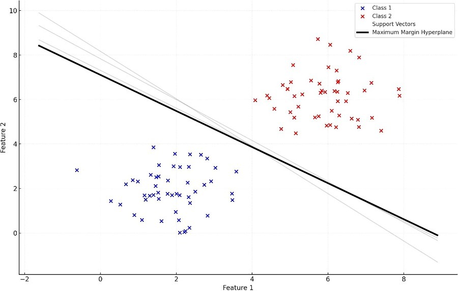
Рисунок 12. Примеры границ решений в разных размерностях: линия в 2D, плоскость в 3D и гиперплоскость в пространстве большей размерности. Такие границы, определяемые SVM, необходимы для оптимального разделения классов и используются, например, для выявления железодефицитного хлороза сои и ржавчины/мучнистой росы пшеницы. SVMможет быть менее эффективна при шуме и нечётких границах (Naik et al., 2017; Xie et al., 2016; Cen et al., 2017).
4.2.1 Метод опорных векторов
SVM — метод обучения с учителем для классификации неизвестных объектов по размеченной выборке. PCA может уменьшить исходный набор, например более чем на 90%, до двух компонент, отображаемых на координатной плоскости. Линия границы оптимально разделяет известные классы, например стресс/здоровое состояние (рисунок 12; Cervantes et al., 2020). В трёх измерениях граница — плоскость, в больших — гиперплоскость. Хотя базовая SVM линейна, ядерные функции создают нелинейные границы; для нескольких классов строят несколько границ. После обучения они классифицируют новые объекты. Несмотря на сотни измерений спектра и миллионы пикселей изображения, снижение размерности часто оставляет компактный набор, например 11 PC, достаточный для классификации. SVM широко применялась в аграрных исследованиях (Naik et al., 2017; Cen et al., 2017; Xie et al., 2016) и особенно подходит малым спектральным выборкам после PCA.
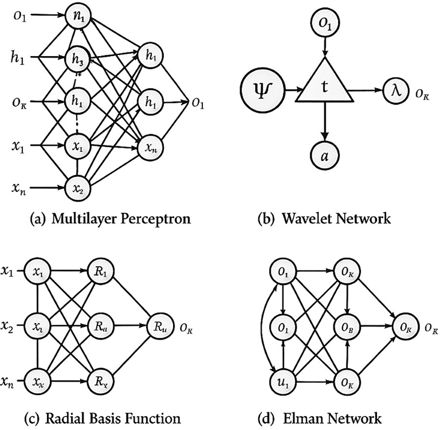
Рисунок 13. Четыре типа искусственных нейронных сетей: (a) многослойный перцептрон с входами xᵢ, скрытыми нейронами hᵢ, выходами Oᵢ и весами wᵢ; (b) вейвлет-сеть с функцией ψ, коэффициентом сдвига tᵢ и масштаба λ; (c) сеть радиально-базисных функций Rᵢ; (d) сеть Элмана с рекуррентными связями, скрытым и выходным слоями и контекстными блоками uᵢ.
4.2.2 Искусственная нейронная сеть
ИНС имитирует структуру и функционирование биологической нервной сети (Krenker et al., 2011). Соединённые искусственные нейроны получают множество входов и выдают результат с учётом их весовой значимости (Sladojevic et al., 2016). ИНС применялись для диагностики прикорневой гнили пшеницы (Humpal et al., 2020), мучнистой росы и мягкой гнили кабачка (Pineda et al., 2017), биотических стрессов граната (Golhani et al., 2019), оранжевой пятнистости масличной пальмы (Dhakate and Ingole, 2015). Они не требуют глубокого знания предметной области, но подвержены переобучению и могут требовать много времени вычислений (Tu, 1996). Типы архитектур показаны на рисунке 13 (Elsheikh et al., 2019). В крупных задачах с изображениями базовые ИНС обычно уступают CNN, но конкурентоспособны на малых табличных/спектральных признаках.
4.2.3 Глубокое обучение
Глубокое обучение — подраздел ML, применяющий многослойные ИНС; «глубина» означает число слоёв. CNN выполняет свёртки над входом и широко применяется в сельском хозяйстве (Jin et al., 2017): для вирусной пестролепестности тюльпана (Polder et al., 2019b), вируса картофеля Y (Polder et al., 2019a), тяжести чёрной гнили яблони (Wang et al., 2017), биотических стрессов огурца (Ma et al., 2018) и кофе (Esgario et al., 2020). U-Net и U-Net++ повысили точность сегментации болезней листьев и картирования стресса полога (Farooqui et al., 2023; Saleem et al., 2020; Zhang et al., 2023b). Распространены предобученные VGG, AlexNet, GoogLeNet и ResNet (Mohanty et al., 2016; Zhang et al., 2018; Ferentinos, 2018; Ma et al., 2018), обучаемые на Diseased Wheat Database и PlantVillage (Lu et al., 2017a; Saleem et al., 2019).
Преимущество глубоких моделей — работа с сырыми данными без тяжёлых ручных стадий цветовых преобразований, снижения размерности, сегментации и выделения признаков (Brahimi et al., 2017); признаки обучаются автоматически (LeCun et al., 2015). Основной недостаток — необходимость сотен или тысяч изображений (Ferentinos, 2018; Dyrmann et al., 2016; Lu et al., 2017b). Трансформеры исследуются благодаря дальним зависимостям и мультимодальному слиянию (Li et al., 2023), особенно для RGB + тепловых + флуоресцентных/HSI входов, но требуют больше вычислений. SVM/RF сильны на малых спектральных данных; ИНС гибки, но переобучаются; CNN/трансформеры наиболее точны на сложных изображениях, но дороги и менее интерпретируемы; ансамбли/гибриды занимают промежуточное положение. Выбор зависит от объёма и типа данных, ресурсов и назначения.
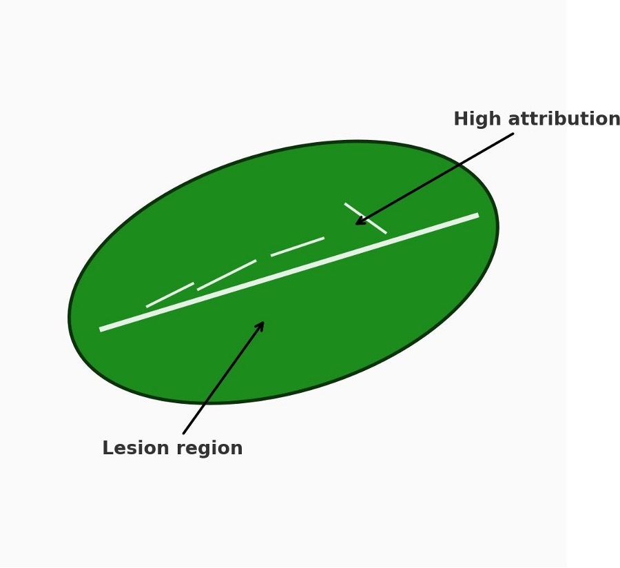
Рисунок 14. Пример визуализации внимания/атрибуции, например Grad-CAM, на поражённых областях листа для повышения интерпретируемости; связан с разделом 4.2.4.
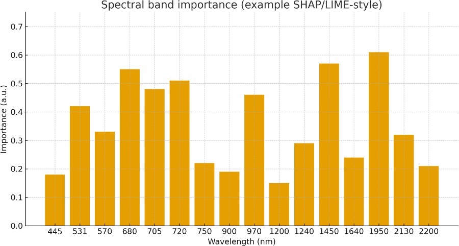
Рисунок 15. Пример атрибуции спектральных признаков в стиле SHAP/LIME, показывающий важность полос при классификации стресса.
4.2.4 Критические аспекты моделей ML и эталонного тестирования
Несмотря на прогресс, надёжность и переносимость моделей ограничены рядом факторов.
4.2.4.1 Ограниченность данных и дисбаланс классов
Большинство работ обучает модели на малых наборах одной культуры в контролируемых условиях, что ограничивает перенос между видами, участками, сезонами и датчиками. Часто здоровый класс значительно больше стрессового, поэтому редкие состояния распознаются хуже. Недостаточно используются стратифицированная выборка, стоимостно-чувствительное обучение, focal loss, калиброванные пороги и синтетическое увеличение меньшинства с учётом риска сдвига распределения. Перенос обучения, адаптация домена и активное обучение уменьшают нагрузку разметки, но применяются неравномерно.
4.2.4.2 Переобучение и утечка при оценке
ИНС и глубокие модели с большим числом параметров особенно склонны переобучаться на ограниченных однородных данных: высокие внутренние показатели ухудшаются в реальных условиях. Помогают weight decay, dropout, аугментация, ранняя остановка и ансамбли, но критична схема оценки. Предпочтительны вложенная перекрёстная проверка, разделение по растениям/делянкам/полям без утечки и внешняя валидация между участками, годами и датчиками, а не случайное разбиение.
4.2.4.3 Интерпретируемость и поддержка решений
«Чёрный ящик» CNN и трансформеров ограничивает доверие. SHAP, LIME и Grad-CAM показывают, какие признаки, полосы или области изображения определяют прогноз, позволяя агроному проверить физиологическую правдоподобность и обнаружить ложные связи, например с почвой или метками. Постфактум-объяснения могут быть нестабильны, поэтому нужны проверки возмущением, оценка вариабельности объяснений и связь атрибуций с физиологическими механизмами.
4.2.4.4 Сопоставимость и эталонные наборы
Сравнение работ затрудняют разные наборы, обработка и метрики. Нужны общественные бенчмарки с фиксированными train/validation/test-разбиениями, версиями и едиными показателями: accuracy, precision, recall, F1, ROC-AUC, PR-AUC для дисбаланса, Brier score и калибровочные кривые. Нужны общие мультимодальные наборы RGB, тепловых, флуоресцентных и HSI-данных и задачи междоменного переноса. Публикация матриц ошибок, доверительных интервалов, кода, конвейеров и «карточек» моделей/данных повысит воспроизводимость.
4.3 Протоколы валидации, обобщения и оценки
Следует различать внутреннюю проверку, например k-кратную внутри набора, и внешнее тестирование в независимых условиях — другой участок, сезон, сорт или сенсор. Внутренняя проверка оценивает работу в пределах распределения, внешняя — реальное обобщение при изменении освещения, структуры полога, фенологии и фона. Рекомендуется сообщать обе и явно указывать разбиение по участкам, видам и датчикам.
Используйте стратифицированную k-кратную проверку без утечки одного растения или делянки между фолдами; при временной корреляции — блочную или групповую CV. Оставляйте минимум один участок или сезон как внешний тест. Сообщайте показатели по участкам/видам и макроусреднение. Для редких ранних симптомов применяйте веса классов, ресэмплинг, focal loss и настройку порога по precision–recall; всегда указывайте распределение классов и стратегию. Одной accuracy недостаточно: нужны precision, recall, macro/micro-F1, PR-AUC и матрицы ошибок; для тяжести как регрессии — R², RMSE/MAE и анализ Бланда–Альтмана.
Перенос обучения от крупных визуальных моделей и лёгкие трансформеры повышают эффективность данных и устойчивость к междоменной вариабельности. Работы по трансформерам (Li et al., 2023), полуконтролируемому обучению при малом числе меток (Huang et al., 2023) и эффективным CNN/ViT для ограниченных ресурсов (Patel et al., 2024a; Debnathand Basu, 2025) показывают выгоды, переносимые в поле. Постоянство цвета, аугментация и аграрно-ориентированная нормализация повышают устойчивость между устройствами и средами (Li et al., 2025a). Модели следует оценивать при (i) предобучении на общих корпусах растений/стресса, (ii) тонкой настройке на целевых культурах/участках и (iii) исключении одного участка по очереди. Полуавтоматическая и масштабируемая разметка помогает сокращать доменный разрыв (Tang etal., 2023b; Mi et al., 2020b).
5 Заключительные замечания
Для различения биотических и абиотических стрессоров, особенно болезней, используются разные оптические датчики и алгоритмы. Машинное обучение регулярно обрабатывает изображения, прежде всего RGB, тогда как в спектроскопии чаще применяются классические статистические методы; однако интеграция ML в спектральный анализ быстро растёт. Настоящий обзор выходит за рамки описания и предлагает сравнительную матрицу решений (таблицы 4a, 4b), оценивающую модальности и алгоритмы по эффективности, стоимости, мобильности и масштабируемости. Рисунки и таблицы встроены в повествование: рисунки 1–4 задают спектральный и аппаратный контекст, таблицы 1 и 2 объединяют длины волн и индексы, а последующие таблицы связывают выбор датчика с алгоритмом и результатами.
Многие исследования применимы лишь к ограниченному числу видов. Значительные различия отражательных свойств листьев затрудняют общие выводы для разных культур и сред. Вероятно, будущая диагностика будет стремиться к универсальности вместо видоспецифичности, но необходимо установить определяющие признаки и параметры. Разрыв межвидового обобщения обосновывает потребность в бенчмарках и внешней валидации, подчёркнутую в разделе 4.2.4 и, где возможно, таблице 6.
Смартфонная RGB- и флуоресцентная визуализация и термография могут масштабироваться до полога в открытой и контролируемой среде. Новые системы стали качественнее и компактнее. Оптическое разрешение смартфонов сопоставимо с отдельными камерами, чувствительность и встроенные вычисления позволяют анализировать данные на устройстве. Облако и удалённое управление файлами поддерживают тяжёлые задачи; оптический зум 2×–4× дополнительно расширяет полевое применение. Однако различение отдельных стрессов, особенно конкретных дефицитов питания, остаётся трудным: рост чувствительности повышает восприимчивость к шуму среды. Сегментация и ML-конвейеры помогают отделять шум. Практически это означает RGB/мультиспектральные + RF/SVM системы для широкого охвата и переход к HSI + CNN/трансформерам в ценных культурах или исследованиях, а также использование индексов таблицы 2 на устройстве.
5.1 Ограничения и трудности
До массового применения остаются препятствия. Высокая стоимость сенсоров, дрейф калибровки и отсутствие стандартных протоколов мешают согласованности систем. Изменения света, влажности и температуры создают сильный полевой шум и уменьшают устойчивость моделей. Малые или несбалансированные обучающие наборы ограничивают перенос между видами и стрессами. Глубокие модели требуют больших размеченных массивов и вычислений, а их «чёрный ящик» затрудняет агрономические решения. Эти факторы объясняют разброс результатов таблицы 6. Необходимы единая отчётность о разбиениях, контроле освещения, журналах калибровки и метриках по классам — precision/recall/F1, ROC-AUC/PR-AUC и калибровке.
5.2 Проблемы внедрения и этические аспекты
5.2.1 Сложности слияния данных
Объединение HSI, мультиспектральных, тепловых, флуоресцентных и RGB-данных сталкивается с несоответствием пространственного разрешения, спектрального покрытия, частоты кадров, освещения и геометрии наблюдения. Ошибки совмещения и радиометрический дрейф нивелируют преимущества. Практические меры: (i) совмещение по реперным меткам/позе БПЛА; (ii) радиометрическая и световая нормализация по панелям и журналам облучённости; (iii) стандартизированные метаданные — сенсор, оптика, высота, угол солнца; (iv) архитектуры по условиям: раннее слияние данных при хорошем выравнивании, слияние признаков при небольшом рассогласовании, позднее слияние решений при неоднородных или отсутствующих модальностях. Следует сообщать абляции, калибровку неопределённости и устойчивость к пропускам. Внедрение разумно начинать с индексов таблицы 2 и усложнять слияние лишь при устойчивом выигрыше в точности или ранности.
5.2.2 Экономическая осуществимость
Высококлассные HSI и крупные глубокие модели чувствительны, но требуют значительных капитальных затрат на оборудование и операционных затрат на калибровку, обслуживание, разметку и вычисления. Смартфон + насадка, ручной мультиспектральный прибор или БПЛА с RGB/мультиспектром имеют привлекательную совокупную стоимость при целевых индексах и компактных моделях. Практическая формула:
Чистая выгода = (сохранённый урожай × цена) + экономия ресурсов − капитальные затраты − операционные затраты − стоимость времени обучения.
Период амортизации должен быть задан. Кооперативы и модель «зондирование как услуга» распределяют затраты; вычисления на краю/устройстве уменьшают расходы на облако и зависимость от связи. Вместе с точностью следует сообщать стоимость на гектар, время до предупреждения и экономию труда.
5.2.3 Барьеры принятия
Помимо цены, внедрение ограничивают обучение персонала, удобство, связь и интеграция. Полевые инструменты должны предоставлять: (i) пошаговые процессы с подсказками калибровки и проверкой качества; (ii) локализованный интерфейс и режимы для пользователей с низкой грамотностью; (iii) автономную работу на устройстве с необязательной облачной синхронизацией; (iv) совместимость с системами управления хозяйством, DSS и IoT. Важны консультационные программы и обслуживание поставщика. Для малых хозяйств начальной ступенью служат RGB/мультиспектр + RF/SVM, а для ценных культур и исследований — HSI + CNN/трансформеры.
5.2.4 Регуляторные и этические аспекты
ИИ-диагностика ставит вопросы владения данными, конфиденциальности, согласия, безопасности и ответственности, когда рекомендации определяют орошение или применение пестицидов. Надлежащая практика включает:
· Управление: ясные правила использования данных; по возможности обработка на устройстве; шифрование; добровольная передача данных; федеративное обучение при проблемах приватности или связи, чтобы обновлять модель без централизации сырых данных.
· Прозрачность и ответственность: участие человека в критически значимых действиях; объяснения, связывающие признаки модели с физиологией; журналы рекомендаций и действий.
· Справедливый доступ: недопущение роста цифрового неравенства; дешёвые тарифы, субсидии или кооперативы; открытые стандарты и совместимость против привязки к поставщику.
· Экологическая ответственность: жизненный цикл устройств, ремонтопригодность и электронные отходы; энергозатраты обучения и вывода; предпочтение лёгких моделей.
5.3 Приоритетная дорожная карта исследований на 5–10 лет
Для надёжного полевого внедрения необходимо закрыть несколько пробелов. Дорожная карта отдаёт приоритет краткосрочной стандартизации и эффективности данных, среднесрочному слиянию и системной интеграции, долгосрочной масштабируемости и равноправию. Этапы следует оценивать по сочетаниям модальностей и алгоритмов в таблицах 4a, 4b, полевым сценариям таблицы 3 и результатам таблицы 6.
5.3.1 Краткосрочный этап, 1–3 года
· Калибровка и протоколы: публиковать минимальные контрольные списки регистрации и калибровки: световые эталоны, журналы облучённости, излучательная способность, оптика/высота/солнечная геометрия, аудиторский след. Предоставлять эталонные панели и открытые скрипты.
· Наборы и метки: выпускать размеченные открытые данные разных видов, участков, сезонов, устройств и стрессоров со стандартными метаданными и сбалансированными разбиениями для редких классов.
· Лёгкие модели: разрабатывать модели для смартфона/края с квантованием, прореживанием и дистилляцией; целевые показатели — менее 20 МБ, вывод менее 200 мс и экономный расход батареи.
· Корректная оценка: исключать утечку по делянкам/полям/годам, использовать внешние тесты, сообщать precision/recall/F1, ROC-AUC/PR-AUC, Brier и кривые надёжности, время до предупреждения, абляции, неопределённость и устойчивость к пропускам.
5.3.2 Среднесрочный этап, 3–5 лет
· Мультимодальное слияние: развивать раннее, признаковое и решающее слияние HSI, тепловых, флуоресцентных и RGB-данных согласно ограничениям выравнивания и доступности; публиковать эталонные конвейеры и предобученные энкодеры.
· Объяснимая поддержка решений: сочетать точные модели с физиологически связанными объяснениями полос/областей для проверки агрономом; стандартизировать устойчивость объяснений и пороги участия человека.
· Интеграция IoT/DSS: связывать зондирование с DSS и IoT для дифференцированного опрыскивания и фертигации, с журналами, ролями и возможностью отката.
· Эффективное обучение: перенос, само- и полуконтролируемое, активное и слабое обучение; целевой показатель — сокращение разметки минимум на 30% без потери точности на двух новых участках.
· Стоимость/эффективность: вместе с точностью сообщать стоимость на гектар, экономию труда и время работоспособности при полевой изменчивости.
5.3.3 Долгосрочный этап, 5–10 лет
· Универсальная недорогая диагностика: модели, обобщающиеся между видами, регионами и устройствами за счёт адаптации домена и метаобучения; управление дрейфом непрерывным обучением и периодической перекалибровкой.
· Масштабные платформы: зрелые смартфонные и БПЛА-системы мониторинга больших площадей с выводом на краю, эпизодической облачной синхронизацией и кооперативными/сервисными бизнес-моделями.
· Экосистемы, сохраняющие приватность: вывод на устройстве и федеративное обучение, при котором сырые данные остаются локальными.
· Стандарты и управление: полевые форматы данных, API, требования безопасности и регулирования, права на данные и ответственность, подготовка и сертификация пользователей.
· Показатели устойчивости: влияние жизненного цикла, ремонтопригодность, электронные отходы и энергия вычислений; предпочтение лёгких моделей и долговечного оборудования.
5.3.4 Эталонное тестирование дорожной карты
На каждом этапе следует оценивать цели по сочетаниям модальностей и алгоритмов из таблиц 4a–4b, согласованным со сценариями таблицы 3 — от дешёвого обследования до точного фенотипирования, — и обобщать результаты по таблице 6. Помимо точности нужны калибровка, время до предупреждения, стоимость на гектар и устойчивость к отсутствующей модальности.
5.4 Практическое применение
Перенос зондирования и аналитики в повседневную агрономию требует надёжных, доступных и простых путей. Смартфонные системы и недорогие переносные датчики расширяют доступ в условиях ограниченных ресурсов, обеспечивая наблюдение болезней в реальном времени, раннее предупреждение о засухе и дефиците питания и локализованное управление. Связь диагностики с DSS и IoT замыкает цикл от обнаружения к действию: дифференцированному опрыскиванию, фертигации/орошению и приоритетному обследованию по картам риска. Критичны автономный вывод, минимальные процедуры калибровки и совместимые стандарты RGB, тепловых, флуоресцентных и спектральных данных. Партнёрства производителей, консультационных служб и ag-tech-компаний ускоряют распространение, согласуя потребности с возможностями и обслуживанием. Производитель может начать с RGB/мультиспектра + RF/SVM и индексных признаков, затем перейти к HSI и трансформерам/CNN, если нужна точность или ранняя биохимическая чувствительность; тепловые/флуоресцентные потоки повышают надёжность предупреждения.
Интеграция RGB, NIR и SWIR с машинным обучением переводит диагностику от видоспецифичных экспериментов к масштабируемым полевым инструментам. Стоимость, калибровка, средовая изменчивость и нехватка данных сохраняются, но мультимодальное слияние, объяснимый ИИ, адаптация домена и смартфоны формируют практическую дорожную карту. Следующий этап требует: (i) стандартной калибровки слияния для согласованности датчиков; (ii) открытых размеченных мультимодальных наборов с межвидовой и межсредовой вариабельностью; (iii) полевых протоколов съёмки, обработки и тестирования. Это необходимо для обобщения и крупномасштабного внедрения. Встраивание в DSS/IoT свяжет зондирование с действиями в реальном времени и создаст надёжные, интерпретируемые и справедливые системы устойчивого сельского хозяйства. Рисунки 1–15 и таблицы 1–6 образуют единую структуру от физики и датчиков через индексы и модели к практическим воздействиям.
Заявление о доступности данных
Исходные материалы исследования включены в статью и дополнительные материалы. Дополнительные запросы можно направлять автору для корреспонденции.
Вклад авторов
MS: концепция, курирование данных, формальный анализ, исследование, методология, программное обеспечение, валидация, визуализация, подготовка исходного текста, рецензирование и редактирование. SK: концепция, курирование данных, формальный анализ, исследование, методология, программное обеспечение, валидация, визуализация, подготовка исходного текста, рецензирование и редактирование. AQ: привлечение финансирования, администрирование проекта, ресурсы, руководство, валидация, подготовка исходного текста, рецензирование и редактирование, методология. HA:ресурсы, руководство, валидация, подготовка исходного текста, рецензирование и редактирование, формальный анализ, методология.
Финансирование
Авторы заявляют, что получили финансовую поддержку исследования и/или публикации статьи. Они благодарят Deanship of Research and Graduate Studies King Khalid University за финансирование в рамках Large Research Project, грант RGP2/283/46.
Благодарности
Автор для корреспонденции благодарит Deanship of Research and Graduate Studies King Khalid University за щедрую поддержку. MS также благодарит кафедру компьютерных наук CECOS University of IT and Emerging Sciences, Пешавар, Пакистан; SK — кафедру компьютерных наук и информационных технологий University of Lahore, Лахор, Пакистан.
Конфликт интересов
Авторы заявляют, что исследование проводилось при отсутствии коммерческих или финансовых отношений, которые могли бы быть истолкованы как потенциальный конфликт интересов.
Заявление об использовании генеративного ИИ
Авторы заявляют, что при создании рукописи генеративный ИИ не использовался.
Любой альтернативный текст, сопровождающий рисунки статьи, был создан Frontiers при поддержке искусственного интеллекта; были предприняты разумные усилия для обеспечения точности, включая проверку авторами, где это было возможно. При обнаружении проблем просьба связаться с издательством.
Примечание издателя
Все утверждения статьи принадлежат исключительно авторам и не обязательно отражают позицию организаций, издателя, редакторов или рецензентов. Любой продукт, оцениваемый в статье, и заявления его производителя не гарантируются и не одобряются издателем.
Список литературы
Adhikari, R., Li, C., Kalbaugh, K., and Nemali, K. (2020). A low-cost smartphone controlled sensor based on image analysis for estimating whole-plant tissue nitrogen (N) content in floriculture crops. Comput. Electron. Agric. 169, 105173. doi: 10.1016/ j.compag.2020.105173
Al Bashish, D., Braik, M., and Bani-Ahmad, S. (2010). “A framework for detection and classification of plant leaf and stem diseases,” in Proceedings of the 2010 international conference on signal and image processing(Chennai, India: IEEE), 113–118.
Allen, W., and Richardson, A. (1968). Interaction of light with a plant canopy. J. Optical Soc. America 58, 1023. doi: 10.1364/JOSA.58.001023
Ammar, M., Hussain, A., and Khan, B. (2024). Enhancing plant disease detection accuracy with YOLOv8- Seg and deep feature fusion. Artif. Intell. Agric. 9, 56–68. doi: 10.1016/j.aiia.2024.03.004
Andolfi, A., Cimmino, A., Evidente, A., Iannaccone, M., Capparelli, R., Mugnai, L., et al. (2009). A new flow cytometry technique to identify Phaeomoniella chlamydospora exopolysaccharides and study mechanisms of esca grapevine foliar symptoms. Plant Dis. 93, 680–684. doi: 10.1094/PDIS-93-7-0680
Aronov, D., Ben-David, S., Kahana, Z., and Shani, E. (2024). Cropvoice: Fluorescent stress signaling in genetically encoded plants enables remote monitoring of crop health. Nat. Biotechnol. 42, 567–574. doi: 10.1038/s41587-024-02058-9
Ashourloo, D., Mobasheri, M., and Huete, A. (2014). Developing two spectral disease indices for detection of wheat leaf rust (Puccinia triticina). Remote Sens. 6, 4723–4740. doi: 10.3390/rs6054723
Bai, G., Jenkins, S., Yuan, W., Graef, G. L., and Ge, Y. (2018). Field-based scoring of soybean iron deficiency chlorosis using rgb imaging and statistical learning. Front. Plant Sci. 9. doi: 10.3389/fpls.2018.01002
Balasundram, S., Golhani, K., Shamshiri, R., and Vadamalai, G. (2020). “Precision agriculture technologies for management of plant diseases,” in Plant disease management strategies for sustainable agriculture through traditional and modern approaches of sustainability in plant and crop protection. Eds. I. Ul Haq and S. Ijaz (Basel, SwitzerlandLawson : Cham: Springer), 259–278, 13. doi: 10.1007/978-3-030-35955-312
Banerjee, K., Krishnan, P., and Das, B. (2020). Thermal imaging and multivariate techniques for characterizing and screening wheat genotypes under water stress condition. Ecol. Indic. 119, 106829. doi: 10.1016/j.ecolind.2020.106829
Bebronne, R., Carlier, A., Meurs, R., Leemans, V., Vermeulen, P., Dumont, B., et al. (2020). In-field proximal sensing of septoria tritici blotch, stripe rust, and brown rust in winter wheat by means of reflectance and textural features from multispectral imagery. Biosyst. Eng. 197, 257–269. doi: 10.1016/j.biosystemseng.2020.02.016
Behmann, J., Acebron, K., Emin, D., Bennertz, S., Matsubara, S., Thomas, S., et al. (2018). Specim IQ: Evaluation of a new, miniaturized handheld hyperspectral camera and its application for plant phenotyping and disease detection. Sensors 18, 441. doi: 10.3390/s18020441
Berdugo, C., Zito, R., Paulus, S., and Mahlein, A.-K. (2014). Fusion of sensor data for the detection and differentiation of plant diseases in cucumber. Plant Pathol. 63, 1344– 1356. doi: 10.1111/ppa.12176
Bone, R., Lee, D., and Norman, J. (1985). Epidermal cells functioning as lenses in leaves of tropical rain-forest shade plants. Appl. Optics 24, 1408. doi: 10.1364/ AO.24.001408
Bornman, J., and Vogelmann, T. (1991). Effect of UV-B radiation on leaf optical properties measured with fibre optics. J. Exp. Bot. 42, 547–554. doi: 10.1093/jxb/ 42.5.547
Brahimi, M., Boukhalfa, K., and Moussaoui, A. (2017). Deep learning for tomato diseases: Classification and symptoms visualization. Appl. Artif. Intell. 31, 299–315. doi: 10.1080/08839514.2017.290263
Brambilla, M. (2020). Application of a low-cost RGB sensor to detect basil (Ocimum basilicum L.) nutritional status at pilot scale level. Precis. Agric. 20. doi: 10.1007/s11119- 020-09788-0
Brooks, M., and Niyogi, K. (2011). “Use of a pulse-amplitude modulated chlorophyll fluorometer to study the efficiency of photosynthesis in Arabidopsis plants,in Chloroplast Research in Arabidopsis, vol. 775Ed. R. P. Jarvis (Humana Press, Totowa, NJ), 299–310. of Methods in Molecular Biology.
Burling, K., Hunsche, M., and Noga, G. (2011). Use of blue–green and chlorophyll fluorescence measurements for differentiation between nitrogen deficiency and pathogen infection in winter wheat. J. Plant Physiol. 168, 1641–1648. doi: 10.1016/ j.jplph.2011.04.004
Buschmann, C., and Lichtenthaler, H. (1998). Principles and characteristics of multi- colour fluorescence imaging of plants. J. Plant Physiol. 152, 297–314. doi: 10.1016/ S0176-1617(98)80276–7
Buster, M., Simpfendorfer, S., Guppy, C., Sissons, M., Tighe, M. K., and Flavel, R. J. (2023). Remote detection of Fusarium crown rot in broadacre bread wheat and durum wheat through use of aerial imagery. Crop Pasture Sci. 75, CP23091. doi: 10.1071/CP23091
Cardim Ferreira Lima, M., Krus, A., Valero, C., Barrientos, A., del Cerro, J., and Rolda´n-Go´mez, J. J. (2020). Monitoring plant status and fertilization strategy through multispectral images. Sensors 20, 435. doi: 10.3390/s20020435
Carter, G. (1993). Responses of leaf spectral reflectance to plant stress. Am. J. Bot. 80, 239–243. doi: 10.1002/j.1537-2197.1993.tb13720.x
Casari, R., Paiva, D., Silva, V., Ferreira, T., Souza, J., Oliveira, N., et al. (2019). Using thermography to confirm genotypic variation for drought response in maize. Int. J. Mol. Sci. 20, 2273. doi: 10.3390/ijms20092273
Cen, H., Weng, H., Yao, J., He, M., Lv, J., Hua, S., et al. (2017). Chlorophyll fluorescence imaging uncovers photosynthetic fingerprint of citrus huanglongbing. Front. Plant Sci. 8. doi: 10.3389/fpls.2017.01509
Cervantes, J., Garcia-Lamont, F., Rodr´ıguez-Mazahua, L., and Lopez, A. (2020). A comprehensive survey on support vector machine classification: Applications, challenges and trends. Neurocomputing 408, 189–215. doi: 10.1016/j.neucom.2020.03.040
Chen, T., Zhang, J., Chen, Y., Wan, S., and Zhang, L. (2019). Detection of peanut leaf spots disease using canopy hyperspectral reflectance. Comput. Electron. Agric. 156, 677–683. doi: 10.1016/j.compag.2018.12.045
Chen, X., Zhang, L., and Wang, R. (2024). Advancements in smartphone-based RGB imaging for plant stress detection. Comput. Electron. Agric. 210, 107910. doi: 10.1016/ j.compag.2024
Chung, S., Breshears, L., Perea, S., Morrison, C., Betancourt, W., Reynolds, K., et al. (2019). Smartphone- based paper microfluidic particulometry of norovirus from environmental water samples at the single copy level. ACS Omega 4, 11180–11188. doi: 10.1021/acsomega.9b01314
Chung, S., Breshears, L., and Yoon, J.-Y. (2018). Smartphone near infrared monitoring of plant stress. Comput. Electron. Agric. 154, 93–98. doi: 10.1016/ j.compag.2018.08.033
Debnath, A. K., and Basu, S. (2025). MobileplantViT: A lightweight vision transformer based architecture for plant disease classification. Comput. Electron. Agric. 213, 108301. doi: 10.1016/j.compag.2024.108301
de Lima, R., dos Santos, T., Vieira, L., de Lourdes Lu´cio Ferrarese, M., Ferrarese- Filho, O., Donatti, L., et al. (2014). Salt stress alters the cell wall polysaccharides and anatomy of coffee (Coffea arabica l.) leaf cells. Carbohydr. Polymers 112, 686–694. doi: 10.1016/j.carbpol.2014.06.028
Deng, J., Dong, W., Socher, R., Li, L.-J., Li, K., and Fei-Fei, L. (2009). “ImageNet: A large-scale hierarchical image database,” in 2009 IEEE conference on computer vision and pattern recognition (CVPR) (Miami, FL, USA: IEEE Computer Society), 248–255. doi: 10.1109/CVPR.2009.5206848
Dhakate, M., and Ingole, A. B. (2015). “Diagnosis of pomegranate plant diseases using neural network,” in 2015 Fifth National Conference on Computer Vision, Pattern Recognition, Image Processing and Graphics (NCVPRIPG), Patna, India. 1–4 (Piscataway, NJ, USA: IEEE). doi: 10.1109/NCVPRIPG.2015.7490056
Dong, X., Li, Y., Zhang, J., Wang, T., and Chen, Y. (2025). A biohydrogel microneedle patch for leaf-based hydrogen peroxide detection in plants. Nat. Commun. 16, 123. doi: 10.1038/s41467-025-01234-5
Dong, Z., Men, Y., Li, Z., Zou, Q., and Ji, J. (2019). Chlorophyll fluorescence imaging as a tool for analyzing the effects of chilling injury on tomato seedlings. Scientia Hortic. 246, 490–497. doi: 10.1016/j.scienta.2018.11.025
Dyrmann, M., Karstoft, H., and Midtiby, H. (2016). Plant species classification using deep convolutional neural network. Biosyst. Eng. 151, 72–80. doi: 10.1016/ j.biosystemseng.2016.07.004
Ehleringer, J., Bjorkman, O., and Mooney, H. (1976). Leaf pubescence: Effects on absorptance and photosynthesis in a desert shrub. Science 192, 376–377. doi: 10.1126/ science.192.4237.376
Elsheikh, A., Sharshir, S., Abd Elaziz, M., Kabeel, A., Guilan, W., and Haiou, Z. (2019). Modeling of solar energy systems using artificial neural network: A comprehensive review. Solar Energy 180, 622–639. doi: 10.1016/j.solener.2018.11.039
Esgario, J., Krohling, R., and Ventura, J. (2020). Deep learning for classification and severity estimation of coffee leaf biotic stress. Comput. Electron. Agric. 169, 105162. doi: 10.1016/j.compag
Fahrentrapp, J. (2019). Detection of gray mold leaf infections prior to visual symptom appearance using a five-band multispectral sensor. Front. Plant Sci. 10. doi: 10.3389/fpls.2019.00628
Farooqui, N. A., Mishra, A. K., Ray, K., and Mallik, S. (2023). “Leaf disease segmentation using u-net++ architecture,” in International conference on trends in electronics and health informatics (Springer Nature Singapore, Singapore), 769–780. doi: 10.1007/978-981-99-8233-664
Farooqui, N. A., and Ritika, (2019). “A machine learning approach to simulating farmers’ crop choices for drought prone areas,” in Proceedings of ICETIT 2019: emerging trends in information technology (Springer International Publishing, Cham), 472–481. doi: 10.1007/978-3-030-30577-243
Ferentinos, K. (2018). Deep learning models for plant disease detection and diagnosis. Comput. Electron. Agric. 145, 311–318. doi: 10.1016/j.compag.2018.01.007
Food and Agriculture Organization of the United Nations (2017). The future of food and agriculture: trends and challenges (Rome, Italy: Food and Agriculture Organization of the United Nations).
Fourty, T., Baret, F., Jacquemoud, S., Schmuck, G., and Verdebout, J. (1996). Leaf optical properties with explicit description of its biochemical composition: Direct and inverse problems. Remote Sens. Environ. 56, 104–117. doi: 10.1016/0034-4257(95)00236-5
Gamon, J., Pen˜uelas, J., and Field, C. (1992). A narrow-waveband spectral index that tracks diurnal changes in photosynthetic efficiency. Remote Sens. Environ. 41, 35–44. doi: 10.1016/0034-4257(92)90059-S
Garcia, E., Kumar, R., and Lee, S. (2024). Enhanced thermal imaging for early detection of plant water stress using AI-assisted analysis. Sensors 24, 1567. doi: 10.3390/ s24041567
Ghosal, S., Blystone, D., Singh, A., Ganapathysubramanian, B., Singh, A., and Sarkar, S. (2018). An explainable deep machine vision framework for plant stress phenotyping. Proc. Natl. Acad. Sci. U.S.A. 115, 4613–4618. doi: 10.1073/pnas.1714975115
Gitelson, A., Zur, Y., Chivkunova, O., and Merzlyak, M. (2002). Assessing carotenoid content in plant leaves with reflectance spectroscopy. Photochem. Photobiol. 75, 272– 281. doi: 10.1562/0031-8655(2002)075(0272:ACCIPL)2.0.CO;2
Golhani, K., Balasundram, S., Vadamalai, G., and Pradhan, B. (2019). Selection of a spectral index for detection of orange spotting disease in oil palm (Elaeis guineensis Jacq.) using red edge and neural network techniques. J. Indian Soc. Remote Sens. 47, 639–646. doi: 10.1007/s12524-019-01034-9
Gomes, M., da Luz, A., dos Santos, M., do Carmo Pimentel Batitucci, M., Silva, D., and Falqueto, A. (2012). Drought tolerance of passion fruit plants assessed by the OJIP chlorophyll a fluorescence transient. Scientia Hortic. 142, 49–56. doi: 10.1016/ j.scienta.2012.05.032
Grant, L., Daughtry, C., and Vanderbilt, V. (1993). Polarized and specular reflectance variation with leaf surface features. Physiologia Plantarum 88, 1–9. doi: 10.1111/j.1399- 3054.1993.tb01318.x
Habibullah, M., Mohebian, M., Soolanayakanahally, R., Bahar, A., Vail, S., Wahid, K., et al. (2020). Low-cost multispectral sensor array for determining leaf nitrogen status. Nitrogen 1, 67–80. doi: 10.3390/nitrogen1010006
Haralick, R., Shanmugam, K., and Dinstein, I. (1973). Textural features for image classification. IEEE Trans. Systems Man Cybernetics SMC-3, 610–621. doi: 10.1109/ TSMC.1973.4309314
Heim, R., Wright, I., Allen, A., Geedicke, I., and Oldeland, J. (2019). Developing a spectral disease index for myrtle rust (Austropuccinia psidii). Plant Pathol. 68, 738–745. doi: 10.1111/ppa.12993
Huang, W., Guan, Q., Luo, J., Zhang, J., Zhao, J., Liang, D., et al. (2014). New optimized spectral indices for identifying and monitoring winter wheat diseases. IEEE J. Selected Topics Appl. Earth Observations Remote Sens. 7, 2516–2524. doi: 10.1109/ JSTARS.2014.2328634
Huang, Z., Wang, Y., and Liu, Q. (2023). A semi-supervised deep learning framework for crop stress recognition under limited labels. Comput. Electron. Agric. 211, 108035. doi: 10.1016/j.compag.2023.108035
Humpal, J., McCarthy, C., Percy, C., and Thomasson, J. (2020). “Detection of crown rot in wheat utilising near-infrared spectroscopy: Towards remote and robotic sensing,” in Proceedings of the autonomous air and ground sensing systems for agricultural optimization and phenotyping V (Toowoomba, Australia: University of Southern Queensland).
Islam, M., Anh, D., Wahid, K., and Bhowmik, P. (2017). “Detection of potato diseases using image segmentation and multiclass support vector machine,” in 2017 IEEE 30th Canadian Conference on Electrical and Computer Engineering (CCECE). 1–4 (ON, Canada: Windsor).
Jin, K., McCann, M., Froustey, E., and Unser, M. (2017). Deep convolutional neural network for inverse problems in imaging. IEEE Trans. Image Process. 26, 4509–4522. doi: 10.1109/TIP.2017
Jones, H. (1999). Use of infrared thermometry for estimation of stomatal conductance as a possible aid to irrigation scheduling. Agric. For. Meteorology 95, 139–149. doi: 10.1016/S0168-1923(98)00104-0
Kahu, S., Raut, R., and Bhurchandi, K. (2018). Review and evaluation of color spaces for image/video compression. Color Res. Appl. 44, 8–33. doi: 10.1002/col.22224
Kalaji, H., Ba`ba, W., Gediga, K., Goltsev, V., Samborska, I., Cetner, M., et al. (2018). Chlorophyll fluorescence as a tool for nutrient status identification in rapeseed plants. Photosynthesis Res. 136, 329–343. doi: 10.1007/s11120-018-0489-3
Kalaji, H., Oukarroum, A., Alexandrov, V., Kouzmanova, M., Brestic, M., Zivcak, M., et al. (2014). Identification of nutrient deficiency in maize and tomato plants by in vivo chlorophyll a fluorescence measurements. Plant Physiol. Biochem. 81, 16–25. doi: 10.1016/j.plaphy.2014.04.003
Khorsandi, A., Hemmat, A., Mireei, S., Amirfattahi, R., and Ehsanzadeh, P. (2018). Plant temperature- based indices using infrared thermography for detecting water status in sesame under greenhouse conditions. Agric. Water Manage. 204, 222–233. doi: 10.1016/j.agwat.2018.03.020
Kim, J., Lee, H., and Santos, R. (2024). Disease stress index (DSI) for early detection of fungal infections in cereal crops using hyperspectral data. Precis. Agric. 25, 355–368. doi: 10.1007/s11119-023-10015-2
Kitic´, G., Tagarakis, A., Cselyuszka, N., Panic´, M., Birgermajer, S., Sakulski, D., et al. (2019). A new low-cost portab le multispectral optical device for precise plant status assessment. Comput. Electron. Agric. 162, 300–308. doi: 10.1016/j.compag.2019.04.007
Konanz, S., Kocsa´nyi, L., and Buschmann, C. (2014). Advanced multi-color fluorescence imaging system for detection of biotic and abiotic stresses in leaves. Agriculture 4, 79–95. doi: 10.3390/agriculture4010079
Krause, G., and Weis, E. (1984). Chlorophyll fluorescence as a tool in plant physiology: II. interpretation of fluorescence signals. Photosynthesis Res. 5, 139–157. doi: 10.1007/BF00029708
Krenker, A., Bester, J., and Kos, A. (2011). “Introduction to the artificial neural networks,” in Artificial neural networks—Methodological advances and biomedical applications. Ed. K. Suzuki (InTech, Rijeka, Croatia).
Krizhevsky, A., Sutskever, I., and Hinton, G. E. (2017). Imagenet classification with deep convolutional neural networks. Commun. ACM 60, 84–90. doi: 10.1145/3065386
Lang, M., Stiehl, P., Braunova´, Z., and Lichtenthaler, H. K. (1992). Investigations of the blue–green fluorescence emission of plant leaves. Botanica Acta 105, 435–440. doi: 10.1002/j.1438-8677.1992.tb00047.x
Lawson, T., and Vialet-Chabrand, S. (2018). “Chlorophyll fluorescence imaging,” in Photosynthesis of methods in molecular biology, vol. 1770. (UK: Springer), 121–140.
LeCun, Y., Bengio, Y., and Hinton, G. (2015). Deep learning. Nature 521, 436–444. doi: 10.1038/nature14539
Lei, R., Du, Z., Qiu, Y., and Zhu, S. (2016). The detection of hydrogen peroxide involved in plant virus infection by fluorescence spectroscopy. Luminescence 31, 1158– 1165. doi: 10.1002/bio.3190
Lei, H., Kumar, A., and Zhao, M. (2024). A deep hybrid dimensionality reduction approach for multispectral plant disease recognition. Expert Syst. Appl. 237, 122109. doi: 10.1016/j.eswa.2024.122109
Lever, J., Krzywinski, M., and Altman, N. (2017). Principal component analysis. Nat. Methods 14, 641–642. doi: 10.1038/nmeth.4359
Li, H., Pan, Y., Zhang, M., and Wang, C. (2022). Pigment-based hyperspectral imaging for early stress detection in crops: A review. Biosyst. Eng. 217, 60–75. doi: 10.1016/j.biosystemseng.2022.01.009
Li, H., Wang, P., Weber, J., and Gerhards, R. (2017). Early identification of herbicide stress in soybean (Glycine max (l.) merr.) using chlorophyll fluorescence imaging technology. Sensors 18, 21. doi: 10.3390/s18010021
Li, W., Zhang, R., and Chen, M. (2023). Transformer-based models for plant disease classification: A comparative study. IEEE Trans. Artif. Intell. 4, 115–125. doi: 10.1109/ TAI.2023.3245874
Liakos, K., Busato, P., Moshou, D., Pearson, S., and Bochtis, D. (2018). Machine learning in agriculture: A review. Sensors 18, 2674. doi: 10.3390/s18082674
Lichtenthaler, H. K., Gitelson, A., and Lang, M. (1996). Non-destructive determination of chlorophyll content of leaves of a green and an aurea mutant of tobacco by reflectance measurements. J. Plant Physiol. 148, 483–493. doi: 10.1016/ S0176-1617(96)80285-4
Lichtenthaler, H. K., and Rinderle, U. (1988). The role of chlorophyll fluorescence in the detection of stress conditions in plants. CRC Crit. Rev. Analytical Chem. 19, S29– S85. doi: 10.1080/10408348808058088
Liew, O., Chong, P., Li, B., and Asundi, A. (2008). Signature optical cues: Emerging technologies for monitoring plant health. Sensors 8, 3205–3239. doi: 10.3390/s8053205
Ma, J., Du, K., Zheng, F., Zhang, L., Gong, Z., and Sun, Z. (2018). A recognition method for cucumber diseases using leaf symptom images based on deep convolutional neural network. Comput. Electron. Agric. 154, 18–24. doi: 10.1016/ j.compag.2018.08.027
Mahlein, A.-K., Kuska, M. T., Behmann, J., Polder, G., and Walter, A. (2022). Advances in plant disease detection using optical sensors and machine learning: A review. Plant Pathol. 71, 505–520. doi: 10.1111/ppa.13465
Mahlein, A.-K., Rumpf, T., Welke, P., Dehne, H.-W., Plu¨mer, L., Steiner, U., et al. (2013). Development of spectral indices for detecting and identifying plant diseases. Remote Sens. Environ. 128, 21–30. doi: 10.1016/j.rse.2012.09.019
Mastrodimos, N., Lentzou, D., Templalexis, C., Tsitsigiannis, D. I., and Xanthopoulos, G. (2019). Development of thermography methodology for early diagnosis of fungal infection in tab le grapes: The case of Aspergillus carbonarius. Comput. Electron. Agric. 165, 104972. doi: 10.1016/j.compag.2019.104972
Mattupalli, C., Mo, C., Shah, K., and Young, C. (2018). Supervised classification of RGB aerial imagery to evaluate the impact of a root rot disease. Remote Sens. 10, 917. doi: 10.3390/rs10060917
McKenzie, D. B., Hossner, L. R., and Newton, R. J. (1984). Sorghum cultivar evaluation for iron chlorosis resistance by visual scores. J. Plant Nutr. 7, 677–685. doi: 10.1080/01904168409363181
Meng, R., Lv, Z., Yan, J., Chen, G., Zhao, F., Zeng, L., et al. (2020). Development of spectral disease indices for southern corn rust detection and severity classification. Remote Sens. 12, 3233. doi: 10.3390/rs12203233
Mohanty, S. P., Hughes, D. P., and Salathe´, M. (2016). Using deep learning for image-based plant disease detection. Front. Plant Sci. 7. doi: 10.3389/fpls.2016.01419
Moshou, D., Bravo, C., Oberti, R., West, J. S., Ramon, H., Vougioukas, S., et al. (2011). Intelligent multi-sensor system for the detection and treatment of fungal diseases in arable crops. Biosyst. Eng. 10 8, 311–321. doi: 10.1016/ j.biosystemseng.2010.12.001
Murchie, E. H., and Lawson, T. (2013). Chlorophyll fluorescence analysis: A guide to good practice and understanding some new applications. J. Exp. Bot. 64, 3983–3998. doi: 10.1093/jxb/ert221
Naik, H. S., Zhang, J., Lofquist, A., Assefa, T., Sarkar, S., Ackerman, D., et al. (2017). A real-time phenotyping framework using machine learning for plant stress severity rating in soybean. Plant Methods 13, 23. doi: 10.1186/s13007-017-0163-7
Oerke, E.-C., Fro¨ hling, P., and Steiner, U. (2011). Thermographic assessment of scab disease on apple leaves. Precis. Agric. 12, 699–715. doi: 10.1007/s11119-011-9206-6
Ojala, T., Pietika¨inen, M., and Ma¨enpa¨a¨, T. (2002). Multiresolution gray-scale and rotation invariant texture classification with local binary patterns. IEEE Trans. Pattern Anal. Mach. Intell. 24, 971–987. doi: 10.1109/TPAMI.2002.1017623
Osco, L. P., Ramos, A. P. M., Furuya, D. E. G., da Silva Junior, C. A., Marcato Junior, J., de Castro Jorge, L. A., et al. (2020). A convolutional neural network approach for counting and measuring soybean seeds in digital images. Plants 9, 1290. doi: 10.3390/ plants9101290
Pandey, P., Ge, Y., Stoerger, V., and Schnable, J. C. (2017). High throughput in vivo analysis of plant leaf chemical properties using hyperspectral imaging. Front. Plant Sci. 8. doi: 10.3389/fpls.2017.01348
Patel, K. K., Kar, A., Jha, S. N., and Khan, M. A. (2014). Machine vision system: A tool for quality inspection of food and agricultural products. J. Food Sci. Technol. 49, 123–141. doi: 10.1007/s13197-011-0321-4
Patra, B., and Sahoo, S. (2024). Deep learning-based automated assessment of drought stress in potato from uav imagery. Front. Plant Sci. 15. doi: 10.3389/ fpls.2024.1297409
Pe´rez-Bueno, M. L., Pineda, M., and Baro´n, M. (2019). Phenotyping plant responses to biotic stress by chlorophyll fluorescence imaging. Front. Plant Sci. 10. doi: 10.3389/fpls.2019.01135
Pen˜uelas, J., Filella, I., Biel, C., Serrano, L., and Save´, R. (1993). The reflectance at the 950–970 nm region as an indicator of plant water status. Int. J. Remote Sens. 14, 1887–1905. doi: 10.1080/01431169308954010
Pen˜uelas, J., Pin˜ol, J., Ogaya, R., and Filella, I. (1997). Estimation of plant water concentration by the reflectance water index wi (r900/r970). Int. J. Remote Sens. 18, 2869–2875. doi: 10.1080/014311697217396
Petrie, P. R., Wang, Y., Liu, S., Lam, S., Whitty, M. A., and Skewes, M. A. (2019). The accuracy and utility of a low-cost thermal camera and smartphone-based system to assess grapevine water status. Biosyst. Eng. 179, 126–139. doi: 10.1016/ j.biosystemseng.2019.03.015
Pineda, M., Pe´rez-Bueno, M. L., Paredes, V., and Baro´n, M. (2017). Use of multicolour fluorescence imaging for diagnosis of bacterial and fungal infection on zucchini by implementing machine learning. Funct. Plant Biol. 44, 563. doi: 10.1071/ FP16037
Polder, G., van der Heijden, G. W. A. M., van Doorn, J., and Baltissen, T. A. H. (2010). Detection of the tulip breaking virus (tbv) in tulips using optical sensors. Precis. Agric. 11, 397–412. doi: 10.1007/s11119-010-9169-2
Pydipati, R., Burks, T. F., and Lee, W. S. (2006). Identification of citrus disease using color texture features and discriminant analysis. Comput. Electron. Agric. 52, 49–59. doi: 10.1016/j.compag.2006.01.004
Raji, S. N., Subhash, N., Ravi, V., Saravanan, R., Mohanan, C. N., Nita, S., et al. (2015). Detection of mosaic virus disease in cassava plants by sunlight-induced fluorescence imaging: A pilot study for proximal sensing. Int. J. Remote Sens. 36, 2880–2897. doi: 10.1080/01431161.2015.1036090
Ramos-Giraldo, P., Reberg-Horton, C., Locke, A. M., Mirsky, S., and Lobaton, E. (2020). Drought stress detection using low-cost computer vision systems and machine learning techniques. IT Prof. 22, 27–29. doi: 10.1109/MITP.2020.2973597
Rodriguez, M. Z., Comin, C. H., Casanova, D., Bruno, O. M., Amancio, D. R., da Costa, L. F., et al. (2019). Clustering algorithms: A comparative approach. PloS One 14, e0210236. doi: 10.1371/journal.pone
Rosique, F., Navarro, P. J., Ferna´ndez, C., and Padilla, A. (2019). A systematic review of perception system and simulators for autonomous vehicles research. Sensors 19, 648. doi: 10.3390/s19030648
Rouse, J. W., Haas, R. H., Schell, J. A., and Deering, D. W. (1974). “Monitoring vegetation systems in the great plains with erts,” in NASA goddard space flight center, 3rd ERTS-1 symposium (Greenbelt, Maryland, USA: NASA Goddard Space Flight Center), vol. 1. , 9.
Rumpf, T., Mahlein, A.-K., Steiner, U., Oerke, E.-C., Dehne, H.-W., and Plu¨mer, L. (2010). Early detection and classification of plant diseases with support vector machines based on hyperspectral reflectance. Comput. Electron. Agric. 74, 91–99. doi: 10.1016/j.compag.2010.06.009
Saleem, M., Atta, B. M., Ali, Z., and Bilal, M. (2020). Laser-induced fluorescence spectroscopy for early disease detection in grapefruit plants. Photochemical Photobiological Sci. 19, 713–721. doi: 10.1039/C9PP00424A
Saleem, M. H., Potgieter, J., and Mahmood Arif, K. (2019). Plant disease detection and classification by deep learning. Plants 8, 468. doi: 10.3390/plants8110468
Savary, S., Bregaglio, S., Willocquet, L., Gustafson, D., Mason D’Croz, D., Sparks, A., et al. (2017). Crop health and its global impacts on the components of food security. Food Secur. 9, 311–327. doi: 10.1007/s12571-017-0659-1
Savary, S., Willocquet, L., Pethybridge, S. J., Esker, P., McRoberts, N., and Nelson, A. (2019). The global burden of pathogens and pests on major food crops. Nat. Ecol. Evol. 3, 430–439. doi: 10.1038/s41559-018-0793-y
Sawinski, K., Mersmann, S., Robatzek, S., and Bo¨ hmer, M. (2013). Guarding the green: Pathways to stomatal immunity. Mol. Plant-Microbe Interact. 26, 626–632. doi: 10.1094/MPMI-10-12-0259-CR
Shaw, M. W., and Osborne, T. M. (2011). Geographic distribution of plant pathogens in response to climate change: Pathogen distributions and climate. Plant Pathol. 60, 31– 43. doi: 10.1111/j.1365-3059.2010.02346.x
Shrivastava, S., Singh, S. K., and Hooda, D. S. (2015). Color sensing and image processing-based automatic soybean plant foliar disease severity detection and estimation. Multimedia Tools Appl. 74, 11467–11484. doi: 10.1007/s11042-015-2784-x
Singh, A., Ganapathysubramanian, B., Singh, A. K., and Sarkar, S. (2016). Machine learning for high- throughput stress phenotyping in plants. Trends Plant Sci. 21, 110– 124. doi: 10.1016/j.tplants.2015.10.015
Singh, A. K., Misra, A., Chaudhary, J., and Ghosh, A. K. (2023). Hyperspectral imaging: A promising technique for non-destructive plant stress phenotyping. Comput. Electron. Agric. 207, 107721. doi: 10.1016/j.compag.2023.107721
Sladojevic, S., Arsenovic, M., Anderla, A., Culibrk, D., and Stefanovic, D. (2016). Deep neural networks based recognition of plant diseases by leaf image classification. Comput. Intell. Neurosci. 2016, 3289801. doi: 10.1155/2016/3289801
Smith, J. A., Lee, M. T., and Kumar, R. (2021). Impact of climate change on the spread of plant pathogens and pests. Agric. Ecosystem Environ. 310, 107308. doi: 10.1016/j.agee.2021.107308
Strano, M. S., Kim, J., Luo, F., and Huang, M. (2024). Chromatic cof-based ph sensors enable early drought detection in crop plants. Advanced Funct. Materials 34, 2400134. doi: 10.1002/adfm.202400134
Su, W. H., Fennimore, S. A., and Slaughter, D. C. (2019). Fluorescence imaging for rapid monitoring of translocation behaviour of systemic markers in snap beans for automated crop/weed discrimination. Biosyst. Eng. 186, 156–167. doi: 10.1016/ j.biosystemseng.2019.02.003
Sui, X., Zheng, Y., Li, R., Padmanabhan, C., Tian, T., Groth-Helms, D., et al. (2017). Molecular and biological characterization of tomato mottle mosaic virus and development of rt-pcr detection. Plant Dis. 101, 704–711. doi: 10.1094/PDIS-08-16-1144-RE
Sujatha, R., Chatterjee, J. M., Jhanjhi, N. Z., and Brohi, S. N. (2025). Ai-based plant leaf disease detection: A scalable and interpreta ble approach. Sci. Rep. 15, 7331. doi: 10.1038/s41598-025-90921-1
Swarbrick, P. J., Schulze-Lefert, P., and Scholes, J. D. (2006). Metabolic consequences of susceptibility and resistance (race-specific and broad-spectrum) in barley leaves challenged with powdery mildew. Plant Cell Environ. 29, 1061–1076. doi: 10.1111/ j.1365-3040.2006.01513.x
Takayama, K., and Nishina, H. (2009). Chlorophyll fluorescence imaging of the chlorophyll fluorescence induction phenomenon for plant health monitoring. Environ. Control Biol. 47, 101–109. doi: 10.2525/ecb.47.101
Tan, M., and Le, Q. V. (2019). Efficientnet: Rethinking model scaling for convolutional neural networks. arXiv preprint arXiv 1905, 11946. doi: 10.5555/3417384.3417966
Tao, M., Ma, X., Huang, X., Liu, C., Deng, R., Liang, K., et al. (2020). Smartphone- based detection of leaf color levels in rice plants. Comput. Electron. Agric. 173, 105431. doi: 10.1016/j.compag.2020.105431
Tsaftaris, S. A., Minervini, M., and Scharr, H. (2016). Machine learning for plant phenotyping needs image processing. Trends Plant Sci. 21, 989–991. doi: 10.1016/ j.tplants.2016.08.003
Tu, J. V. (1996). Advantages and disadvantages of using artificial neural networks versus logistic regression for predicting medical outcomes. J. Clin. Epidemiol. 49, 1225– 1231. doi: 10.1016/S0895-4356(96)00023-4
Vatamanu, O. A., Frandes, M., Ionescu, M., and Apostol, S. (2013). “Content-based image retrieval using local binary pattern, intensity histogram and color coherence vector,” in 2013 E-health and bioengineering conference (EHB)(Iasi, Romania: American Chemical Society (ACS Publications)), 1–6.
Veys, C., Chatziavgerinos, F., AlSuwaidi, A., Hibbert, J., Hansen, M., Bernotas, G., et al. (2019). Multispectral imaging for presymptomatic analysis of light leaf spot in oilseed rape. Plant Methods 15, 4. doi: 10.1186/s13007-019-0391-1
Veys, C., Hibbert, J., Davis, P., and Grieve, B. (2017). “An ultra-low-cost active multispectral crop diagnostics device,” in 2017 IEEE sensors(Glasgow, UK: Taylor & Francis / Informa UK Ltd.), 1–3. doi: 10.1109/ICSENS.2017.8234178
Vilfan, N., van der Tol, C., Yang, P., Wyber, R., Malenovsky´, Z., Robinson, S. A., et al. (2018). Extending fluspect to simulate xanthophyll driven leaf reflectance dynamics. Remote Sens. Environ. 211, 345–356. doi: 10.1016/j.rse.2018.04.002
Wang, J., Patel, A., and Zhang, X. (2025). Advances in porta ble thermography for precision agriculture applications. Agric. Eng. Today 39, 88–99. doi: 10.1016/ j.ageng.2025.01.005
Wang, G., Sun, Y., and Wang, J. (2017). Automatic image-based plant disease severity estimation using deep learning. Comput. Intell. Neurosci. 2017, 2917536. doi: 10.1155/2017/1720198
Watchareeruetai, ,. U., Noinongyao, P., Wattanapaiboonsuk, C., Khantiviriya, P., and Duangsrisai, S. (2018). “s,” in Identification of plant nutrient deficiencies using convolutional neural network (IEEE, Krabi, Thailand), 1–4.
Wei, F., Zhang, Y., and Liu, Q. (2024). Plantseg-net: A deep learning model for precise plant disease segmentation using advanced color space transformation. IEEE Access 12, 45201–45212. doi: 10.1109/ACCESS.2024.3389402
Wold, S., Esbensen, K., and Geladi, P. (1987). Principal component analysis. Chemometrics Intelligent Lab. Syst. 2, 37–52. doi: 10.1016/0169-7439(87)80084-9
Xie, X., Zhang, X., He, B., Liang, D., Zhang, D., and Huang, L. (2016). “A system for diagnosis of wheat leaf diseases based on android smartphone,” in International symposium on optical measurement technology and instrumentation(Beijing, China: Proceedings of Machine Learning Research (PMLR)).
Yang, W., Yang, C., Hao, Z., Xie, C., and Li, M. (2019). Diagnosis of plant cold damage based on hyperspectral imaging and convolutional neural network. IEEE Access 7, 118239–118248. doi: 10.1109/ACCESS.2019.2936582
Yue, J., Li, Z., Liu, L., and Fu, Z. (2011). Content-based image retrieval using color and texture fused features. Math. Comput. Model. 54, 1121–1127. doi: 10.1016/ j.mcm.2011.03.019
Zhang, J., Naik, H. S., Assefa, T., Sarkar, S., Chowda Reddy, R. V., Singh, A., et al. (2017). Computer vision and machine learning for robust phenotyping in genome-wide studies. Sci. Rep. 7, 44048. doi: 10.1038/srep44048
Zhang, K., Wu, Q., Liu, A., and Meng, X. (2018). Can deep learning identify tomato leaf disease? Adv. Multimedia 2018, 1–10. doi: 10.1155/2018/9092501
Zhou, Z.-H. (2018). A brief introduction to weakly supervised learning. Natl. Sci. Rev. 5, 44–53. doi: 10.1093/nsr/nwx181
Zhu, J., He, W., Yao, J., Yu, Q., Xu, C., Huang, H., et al. (2020). Spectral reflectance characteristics and chlorophyll content estimation model of Quercus aquifolioides leaves at different altitudes in sejila mountain. Appl. Sci. 10, 3636. doi: 10.3390/ app10103636
Zhuang, S., Wang, P., Jiang, B., Li, M., and Gong, Z. (2017). Early detection of water stress in maize based on digital images. Comput. Electron. Agric. 140, 461–468. doi: 10.1016/j.compag.2017.07.007
Zovko, M., Zˇ ibrat, U., Knapicˇ, M., Kovacˇic´, M. B., and Romic´, D. (2019). Hyperspectral remote sensing of grapevine drought stress. Precis. Agric. 20, 335–347. doi: 10.1007/s11119-018-9591–1
Zubler, A. V., and Yoon, J.-Y. (2020). Proximal methods for plant stress detection using optical sensors and machine learning. Biosensors 10, 193. doi: 10.3390/ bios10120193
10 августа / 2026